


Spokespersons
Contact
Dr. Sanja Drakulic
Scientific Coordinator
sanja.drakulic@mdc-berlin.de
Phone: 030 9406-1506
Max-Delbrück-Centrum for Molecular Medicine (MDC)
Robert-Rössle-Str. 10
13092 Berlin, Deutschland
Building 84, Room 1015

New molecular and cellular insights obtained from imaging technologies should be integrated with data science into a coherent picture of tissues, organs and organisms, with a goal of prevention, early interception and treatment of diseases.
To contribute to this goal, the Max Delbrück Center for Molecular Medicine; the Weizmann Institute of Science in Rehovot, Israel; the Humboldt-Universität zu Berlin and the Charité-Universitätsmedizin, Berlin have joined forces to establish iNAMES - MDC-Weizmann Helmholtz International Research School (HIRS) for Imaging and Data Science from the NAno to the MESo.

https://www.mdc-berlin.de/inames#t-joinus!
The mission of iNAMES is the training of outstanding imaging and data scientists in a truly international research school, along with answering some of the most important biomedical and clinical research challenges of our time.
The focus of iNAMES Research School lies in the development of an independent, interdisciplinary research project, supported by an international team of mentors from partner institutions from Germany and Israel.
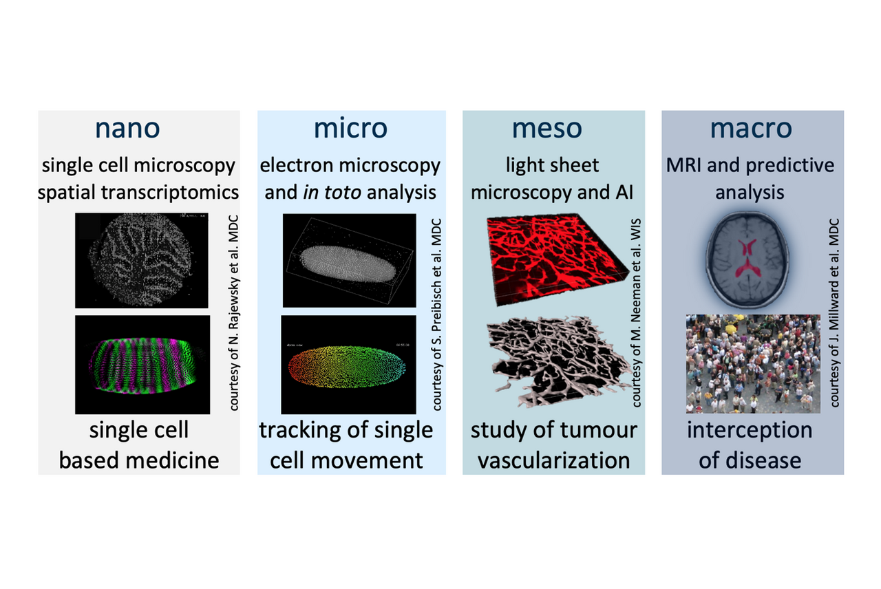
The contribution of participating labs through complementary research studies, models and technologies ensure highly innovative, interdisciplinary projects in the areas of:
The joint effort brings together the understanding of the mechanisms by which diseases originate from defects at the molecular and cellular levels, with developments in techniques that characterize how the cells and their molecular machinery functions at the nanoscopic level, and with emerging non-invasive methods that relate the characterization of tissues and major organ systems at the mesoscopic level.
Examples of cross-domain project topics, combining multiscale imaging with model based analysis, artificial intelligence and machine learning.
The iNAMES program framework includes the following key elements:
We do not only provide our students with a peer network and practical scientific training, but also offer an outstanding opportunity to actively participate in the international scientific community, all ensuring a timely completion of the dissertation. Our overarching goal is to equip our graduates with the critical skills essential for life-long learning, enabling them to successfully compete in any international job market.
The iNAMES PhD researchers are mentored by a tandem of a primary mentor and a co-mentor(s) from partner institutions in Germany and Israel. The mentors support the preparation and implementation of the research project, assist in developing the Individual Development Plan (IDP), introduce to their own professional networks and advise on future plans and career goals.
Besides day-to-day mentors supervision, iNAMES doctoral researchers present their project annually to the Thesis Advisory Committee (TAC), which accompanies them from the first presentation of the research proposal to the preparation of the doctoral thesis. The primary mentor and co-mentor(s), together with two additional independent scientists, form a Thesis Advisory Committee (TAC).
International and cross-disciplinary mobility of scientists between partner sites is of crucial importance for the success of iNAMES. Joint PhD projects are carefully set-up to enable the contribution of participating labs through complementary research, ensuring highly innovative, interdisciplinary projects.
The iNAMES Research School supports short-term stays to learn particular techniques (e.g. for 2-4 weeks) as well as extended stays (e.g. to work on research sub-projects in collaboration with a partner lab, for up to 6 months).
Each iNAMES student is expected to spend at least 2 weeks a year in the partner lab abroad.
To further increase the visibility of iNAMES within the research community, all doctoral researchers are encouraged to network and present their research at international conferences, supported by iNAMES travel and training grants.
iNAMES doctoral researchers are fully funded through an initial 3-year employment contract with the MDC. iNAMES provides funding for a duration of three years, with a possible fourth year needing to be financed by the primary mentor, in accordance with recommendation of the Thesis Advisory Committee (TAC) members. All doctoral researchers are expected to complete their doctoral research and thesis in 4 years.
Salaries are competitive to national standards, according to 65% TVöD 13 (monthly net payment of approximately 1800 EUR). Health and social insurance are included in the contract. The university tuition fees per semester are approximately 350 EUR, and include public transportation ticket.

iNAMES Research School offers advanced, structured and integrated training program with a strong focus on biomedical imaging and data science, in a highly interdisciplinary environment.
Upon completion of the iNAMES training, the graduates:
The iNAMES scientific program is designed to provide doctoral researchers with advanced training on state-of-the-art imaging and data science technologies. A spectrum of iNAMES training activities includes lecture series, student seminars, on-site courses, e-learning courses, hands-on practical seminars, and various scientific events, such as joint international PhD retreats and summer/winter schools.
Every 3rd Wednesday of the month, iNAMES PhD students, associates and guests present their exciting research projects in the area of biomedical imaging and data science.
The "Frontiers in Biomedical Imaging and Image Analysis" series was launched in January 2021. Every 1st Wednesday of the month, excellent scientists working in the area of biomedical imaging and image analysis present their outstanding research to the MDC community and our guests.
A variety of relevant courses is organised for the iNAMES doctoral researchers on site regularly, such as courses in image analysis, programming, statistics, data visualisation and similar. Additionally, doctoral researchers have an opportunity to participate in external courses, supported by the iNAMES training grants. The iNAMES school furthermore organizes info sessions with the MDC technology platforms, hands-on sessions at imaging centers, visits to our clinical partners (Charité and Sheba Medical Center), as well as company visits (e.g. to Siemens, Bruker, Nikon, Zeiss and local start-ups), to foster practical competencies and the entrepreneurship spirit of our doctoral researchers.
To further increase the visibility of iNAMES within the research community, all doctoral researchers are encouraged to network and present their research at international conferences, supported by iNAMES travel and training grants.
Becoming an expert in a specific scientific domain or technology is an important, but not the only aspect of the iNAMES training program. With a growing number of PhDs following careers outside of academia, it is crucial to foster a wider range of personal skills and to introduce relevant career pathways as early as possible.
Our doctoral researchers take responsibility for their professional growth by developing and implementing an Individual Development Plan (IDP). This includes a research plan, training activities, as well as personal skills assessment and progress plan, enabling planning and monitoring of academic and personal development. The IDP is adapted throughout the PhD, particularly as a part of the progress review at the annual TAC meetings.
Peer Mentor Group is introduced as an additional measure to support career development, providing a platform for open discussions on professional and personal skills needs, peer advice on career options, or matters of work/ life balance, and facilitating the integration of iNAMES researchers visiting from partner institutions.
Career Orientation Workshops inform about the available career choices and assist with decisions about the next professional steps.
Technology Transfer Training is essential in preparing for careers both in and outside of academia. The MDC and WIS Technology Transfer Offices offer regular courses and workshops on IPR, patenting, avenues for commercialization of research, translating research into a product, translating basic research into real-world clinical applications, and similar.
Extracurricular and student-organized activities, such as organization of scientific events, teaching at the Life Science Learning Lab “Gläsernes Labor” on the Berlin-Buch campus, participation in public outreach activities during, e.g., “The Long Night of Sciences”, organization of social events, and similar, contribute to the development of the students’ professional skills. These activities are further facilitating the integration of international students, creating a particularly welcoming and lively atmosphere on the campus.
Despite complex circumstances for initiating and driving an international exchange research program in 2020/21, we dived into this task with dedication, passion and optimism. To share our enthusiasm with the community, we organised a virtual kick-off event in September 2021, aiming to introduce the concept of the iNAMES Helmholtz International Research School, its mission and plans for the future, as well as to present the first cohort of our PhD students and their exciting research projects.
As part of the Frontiers in Biomedical Imaging and Image Analysis Lecture Series at the MDC, we were delighted to welcome Wojtek James Goscinski from the Australian National Imaging Facility. He provided a fascinating keynote on Crossroads of Data Science and Imaging: Infrastructure and Applications.
In the iNAMES Orientation session, doctoral researchers are provided with all information regarding the Helmholtz Association and the MDC organizational structure, university regulations, university admission and enrollment, credit points system, obligatory annual Advisory Committee Meetings (TACs), travel details, training grants, training opportunities and more. Within the Orientation session held in September 2021, iNAMES students participated in the Design Thinking workshop, boosting their creativity and collaborative mindset.
iNAMES Orientation Session 2021 | Design Thinking Workshop
The annual iNAMES Symposia, organized alternating between Berlin and Rehovot, gathers a wider scientific and industrial community, stimulating discussion, collaboration and networking between the iNAMES doctoral researchers, faculty, and representatives of industry partners.
More experienced doctoral researchers present their research, providing insight into the state-of-the-art technology developments, while at the same time gaining valuable presentation skills and teaching experience.
A “science café” open to the general public supplements the scientific program of the annual symposia, providing a platform for science communication and outreach.
The 1st iNAMES Symposium "Beyond IMAGEination I." was held at the MDC-BIMSB on November 23-24, 2022. The event gathered 104 participants from 10 institutions, and 25 invited speakers from 6 institutions (EMBL, MPI for Multidisciplinary Sciences, MDC, Charité, HU, WIS) presented their research. In addition, the attendees had the opportunity to enjoy 9 students chalk-talks, 16 poster presentations and 2 networking evening events. The scientific program of the symposium is available here.
Attendees of the first iNAMES Symposiums at MDC-BIMSB.
The 2nd iNAMES Symposium, "Beyond IMAGEination II." will be held at Weizmann Institute of Science in Rehovot, Israel on November 26-27, 2023.
Furthermore, faculty members and doctoral researchers gather at annual summer/ winter schools, dedicated workshops on current advanced topics in imaging and data science.
Trainers: Dr. Elmar Spiegel, Dr. Marina Jiménez Muñoz; Helmholtz Zentrum Munich - German Research Center for Environmental Health, Core Facility Statistical Consulting
Introduction to Statistics and Mixed Models provides a foundation in statistical analysis for scientists and practitioners. The first part covers descriptive statistics, classical statistical tests (t-test, Wilcoxon/Mann-Whitney-U test, ANOVA, etc.), and linear regression and its extensions. The second part deals with repeated measurements, in particular Mixed Models. All methods are explained in an applied setting. By the end of the week, attendees will be able to identify appropriate statistical methods, apply them, and interpret your results.
Trainers: Dr. Elmar Spiegel, Dr. Marina Jiménez Muñoz; Helmholtz Zentrum Munich - German Research Center for Environmental Health, Core Facility Statistical Consulting
Graphics with R covers methods in customizing base R graphics. Additionally, an introduction to ggplot2, an advanced and powerful tool for building graphics with R, is provided. In this course you will learn how to create refined, meaningful graphs in R to visually describe your research outputs. All topics are accompanied with hands-on exercises.
PhD retreats bring together doctoral researchers from partner institutions, providing opportunities to network, exchange ideas and experiences, brainstorm, establish collaborations and form friendships. Doctoral researchers are actively involved in the organization of these meetings, thus developing their organization and project management skills.
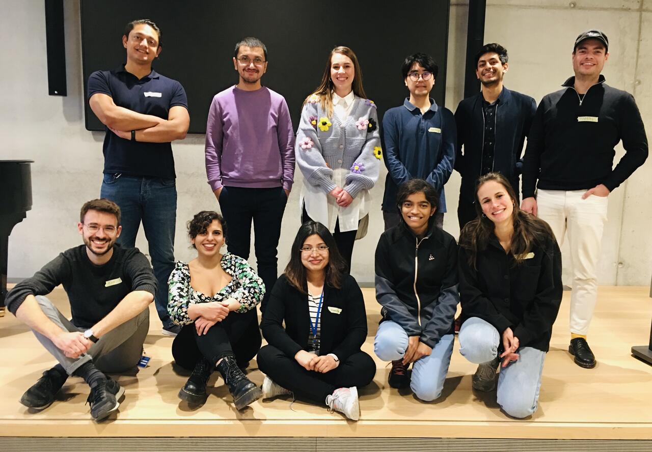
iNAMES doctoral researchers | October 2021
Project: Exploring the molecular mechanism of guanylate-binding proteins at high structural and temporal resolution
Mentor: Oliver Daumke, MDC
Co-mentor: Hagen Hofmann, WIS
iNAMES offers a truly interdisciplinary environment by providing its members with expertise from diverse research fields.
Guanylate-binding proteins are key to cell-autonomous immunity against microbial and viral pathogens. Exploring the molecular mechanism of guanylate-binding proteins requires a set of structural biological approaches in combination with biochemical and cell biological approaches. To this end, we will perform X-ray crystallography, cryo electron microscopy and tomography, single-molecule FRET, and membrane binding and remodeling assays.
Contact: Marius.Weismehl@mdc-berlin.de
Publications:
Weismehl, M., Chu, X., Kutsch, M., Lauterjung, P., Herrmann, C., Kudryashev, M., & Daumke, O. (2024). Structural insights into the activation mechanism of antimicrobial GBP1. The EMBO journal, 43(4), 615–636.
MDC Research Highlight: How a protein fights off bacteria
The best thing about being part of an exchange program is, it creates awareness and acceptance of alternative together with multi-faceted approaches of learning and gaining knowledge.
Project: Development, validation and application of radiofrequency coil technology for ultrahigh field magnetic resonance
Mentor: Thoralf Niendorf, MDC
Co-mentor: Lucio Frydman, WIS / Rita Schmidt, WIS
My project is to develop and implement of radiofrequency (RF) coil technology, which will provide rich opportunities for new ways of non-invasive mapping and probing morphology, function, physiology and metabolism and facilitates explorations into the benefits and challenges of ultrahigh field Magnetic Resonance (UHF-MR) with the ultimate goal to spatially resolve and characterize (patho)physiological processes and biophysical mechanisms to promote a transfer from basic research to clinical science and vice versa.
Publications:
P. Lacour, P. L. Dang, F. R. Heinzel, A. S. Parwani, F. Bähr, A. Kucher, F. Hohendanner, T. Niendorf, F. Rahimi, N. Saha, H. Han, K. Rubarth, M. Sherif, L.-H. Boldt, B. Pieske, F. Blaschke (2022): Magnetic field-induced interactions between phones containing magnets and cardiovascular implantable electronic devices: Flip it to be safe? Heart Rhythm, 19(3):372-380.
N. Saha, J. M. Millward, C. J. J. Herrmann, F. Rahimi, H. Han, P. Lacour, F. Blaschke, T. Niendorf (2023): High-fidelity 3D stray magnetic field mapping of smartphones to address safety considerations with active implantable electronic medical devices. Sensors, 2023, 23(3):1209.
N. Saha, A. Kuehne, J. M. Millward, T. W. Eigentler, L. Starke, S. Waiczies, T. Niendorf (2023): Advanced Radio Frequency Applicators for Thermal Magnetic Resonance Theranostics of Brain Tumors. Cancers, 2023, 15(8):2303.
Contact: Nandita.Saha@mdc-berlin.de
Project: Where is the pain in the brain? Probing the functional neurosignatures of pain at ultrahigh magnetic field strengths
Mentor: Thoralf Niendorf, MDC
Co-mentor: Michal Ramot; WIS
The international collaboration and teamwork not only contribute to achieve a highly enriched project, but also allow me to get to know a different culture.
The project focuses on identifying functional biomarkers for pain to enhance our understanding of pain processing. Despite substantial progress in recent medicine, it is still not understood how pain is processed in the brain. We have no objective biomarkers to assess pain in the patient. Pain cannot be evaluated in comatose or demented patients, and its subjectivity puts an enormous psychological burden on chronic pain patients.
Contact: Igor.Tellez@mdc-berlin.de
Project: Cellular encoding of temperature in the posterior insular cortex
Mentor: James Poulet, MDC
Co-mentor: Ofer Yizhar, WIS
Getting to know other groups and working in different institutes is great for creating networks, which can lead to beautiful collaborations and opportunities.
The ability to perceive temperature helps animals to maintain the homeostasis of body temperature, to avoid noxious temperature that may cause damage, and to identify objects during haptic exploration. My aim in this project is to understand the neural mechanisms of temperature perception in the mouse forelimb system. I plan to use a combination of approaches including two-photon calcium imaging, optogenetic manipulations and behavioural training, to investigate how the thermal cortex represents skin temperature and how this information is translated into thermal perception.
Publications:
M. Vestergaard, M. Carta, G. Güney & J. F. A. Poulet (2023): The cellular coding of temperature in the mammalian cortex, Nature, 614:725–731.
Contact: Gamze.Gueney@mdc-berlin.de
In the news: iNAMES promotes German-Israeli exchange
Project: Computational methods to achieve whole-brain imaging and analyze behavior in animal models to understand Brain-Behavior relationships
Mentor: Kyle Harrington / Hanna Hörnberg / Dagmar Kainmüller, MDC
Co-mentor: Takashi Kawashima, WIS
iNAMES program answers one of the most important aspects of Science – unification: a multi-disciplinary team working towards one solution.
The goal of the project is to develop automatic, real-time algorithms for artifact correction of live fluorescent imaging in the brain of behaving animals. This project especially focuses on cancelling the effect of motion and cerebral blood flow during imaging using a lightsheet microscope and aims to implement such a software module in an open-source microscope control software. We aim to develop novel algorithms to address the existing neural imaging challenges, and to achieve brain-wide voltage imaging, which is one of the technical milestones for neuroscience.
Publications:
R. Barbara, M. Nagathihalli Kantharaju, R. Haruvi, K. Harrington, T. Kawashima (2022): PyZebrascope: An Open-Source Platform for Brain-Wide Neural Activity Imaging in Zebrafish. Frontiers in Cell and Developmental Biology, 10:875044.
L. Finotto, B. Cole, W. Giese, E. Baumann, A. Claeys, M. Vanmechelen, B. Decraene, M. Derweduwe, N. Lakic, G. Shankar, M. Nagathihalli Kantharaju, J. Albrecht, I. Geudens, F. Stanchi, K. Ligon, B. Boeckx, D. Lambrechts, K. Harrington, L. Van Den Bosch, H. Gerhardt (2023): Single‐cell profiling and zebrafish avatars reveal LGALS1 as immunomodulating target in glioblastoma. EMBO Molecular Medicine. 15.
Contact: Madhu.NagathihalliKantharaju@mdc-berlin.de
Project: Developing chimeric human-murine brain slices: a model to study neurodegeneration
Mentor: Volker Siffrin, MDC/Charité
Co-mentor: Michal Schwartz, WIS
The collaborative project, opportunity to be supported and co-mentored during my research project and to acquire shared experiences of involved institutions is the perfect environment to thrive as a young scientist.
Through my clinical work I have seen many patients suffering from neurodegenerative disorders such as Alzheimer`s disease or multiple sclerosis, who are burdened by the limitations of pharmacological agents unable to directly target etiological factors of development of brain degeneration. My project is focused on developing authentic model of neurodegeneration by means of xenografting human pluripotent stem cells from multiple sclerosis patients to develop chimeric brain slices. I will be imaging the cellular and synaptic changes in organ like context by using two photon laser scanning microscopy to visualize complex interplay among different CNS cell populations. These chronic slices might be used in a context of drug treatment, understanding pathological mechanisms of events in cellular and network level.
Publications:
J. Kerkering, B. Muinjonov, K. S. Rosiewicz, S. Diecke, C. Biese, J. Schiweck, C. Chien, D. Zocholl, T. Conrad, F. Paul, M. Alisch, V. Siffrin (2023): iPSC-derived reactive astrocytes from patients with multiple sclerosis protect cocultured neurons in inflammatory conditions. J Clin Invest, 133(13):e164637.
K.S. Rosiewicz, B. Muinjonov, S. Kunz, H. Radbruch, J. Chen, R. Jüttner, J. Kerkering, J. Ucar, T. Crowley, B. Wielockx, F. Paul, M. Alisch, V. Siffrin (2023): HIF prolyl hydroxylase 2/3 deletion disrupts astrocytic integrity and exacerbates neuroinflammation. Glia, 71(8), 2024–2044.
Contact: Bakhrom.Muinjonov@mdc-berlin.de
Project: Quantitative MRI for brain microstructural characterization in multiple sclerosis
Mentor: Friedemann Paul, MDC/Charité / Alexander U. Brandt, University of California, Irvine/ECRC/Charité
Co-mentor: Assaf Tal, WIS
I have always been fascinated by non-invasive imaging techniques to help diagnosis and treatment of clinical pathologies. I find exciting the prospect of developing novel experimental techniques that will eventually find use in a clinical framework.
Autoimmune neuroinflammatory disorders are chronic progressive diseases, leading to disabilities such as visual loss, motor and cognitive impairment. The most prevalent chronic inflammatory disease of the central nervous system is multiple sclerosis. It is characterized by dissemination of white matter and cortical lesions in space and time, driven by disease-specific mechanisms, including inflammation, demyelination, failure to remyelination, axonal damage and other gliopathies. Our aim ist to derive microstructural tissue measurements that are disease and pathology specific, using multiparametric quantitative MRI/MRS methods, and explore tailored analytical approaches for clinical application.
Contact: Henri.Trang@mdc-berlin.de
Project: Neuron-astrocyte coupled lipid clearance dysfunction i n Parkinson’s disease
Mentor: Melissa Birol, MDC
Co-mentor: Phil Selenko, WIS
Although our main location is Berlin, the iNAMES program allows us to get to know the scientific community at the Weizmann institute, to travel there and to learn from its scientists via summer schools and lectures.
In my project, we are aiming to understand how lipid metabolism and more specifically, the interaction between alpha-synuclein with cellular lipoproteins affects its structure, fibril formation, (dis)function and localization in cells. In particular, we are seeking to understand how lipoproteins responsible in coupling the neuron-astrocyte lipid metabolism may affect the aggregation and function of alpha-synuclein. The amyloid fibril formation of alpha-synuclein has been recognized in various neurological diseases including Parkinson's disease. Therefore, understanding how this protein interacts with cellular lipoproteins that couple the neuron-astrocyte lipid metabolism may help us to identify certain parameters that trigger the aggregation and misfunction and thus, lead to the formation of pathogenic alpha-synuclein.
Contact: Paula.SantosOtte@mdc-berlin.de
Project: Renal insight: integrating MRI and machine learning for comprehensive nephrological analysis
Mentor: Thoralf Niendorf, MDC
Co-mentor: Lucio Frydman, WIS
Co-mentor: Erdmann Seeliger, Charité
Science never is a one-(wo)man-show, it is always a team performance. By being part of iNAMES, I can be part of a supportive and familial community which helps me growing and where I can help my colleagues as well.
Kidney diseases are a major health issue, with increasing incidence and an estimated five to ten million deaths per year worldwide. While several biomarkers are currently being investigated for diagnosis of renal diseases, to date clinical point-of-care biomarkers are still lacking for major renal diseases. To address this urgent unmet clinical need, non-invasive imaging may provide markers to inform on the different stages of renal pathophysiology, improve prediction and interception of disease progression and evaluate treatment of renal disease. This project examines the feasibility and reliability of longitudinal monitoring of kidney size in MR images of rats using deep learning approaches. We hypothesize that this approach enables quantification of acute changes in kidney size in experimental setups mimicking realistic clinical scenarios. For this purpose, a deep learning approach for renal segmentation that provides a solution for deforming the kidney shape using a constrained statistical model-based algorithm trained upon a (or few) dataset(s) will be developed. For swift translation of the deep learning approach to clinical data, we will implement a transformative solution that helps to bridge the gap between preclinical and clinical MRI.
Contact: Tobias.Klein@mdc-berlin.de
Publications:
T. Klein, T. Gladytz, J. M. Millward, K. Cantow, L. Hummel, E. Seeliger, S. Waiczies, C. Lippert, T. Niendorf (2023): Dynamic Parametric MRI and Deep Learning: Unveiling Renal Pathophysiology through Accurate Kidney Size Quantification. NMR in Biomedicine, e5057.
Project: Molecular characterization of protein tethers required for touch sensation
Mentor: Gary Lewin, MDC
Co-mentor: Mike Fainzilber, WIS
The key word is networking. iNAMES gives PhD students a great opportunity to meet people and network with them.
At the MDC, I am investigating touch at molecular level. It is striking how little we know about touch, compared to other senses. In the Lewin lab, we are working on a model to explain how the receptors in the skin can transform mechanical forces (i.e. tactile stimuli) into electrical signals, a process known as “mechanotransduction”. This is the first step in touch perception and implies the presence of special ion channels able to be gated by mechanical stimuli. Our model implies that such channels are physically connected to the extracellular matrix by means of large protein tethers and that the stretching of these tethers by mechanical forces opens the channels. My main goal is to visualize these tethers and unveil their molecular identity. Understanding how molecules assemble to function in a mechanotransduction complex will open up avenues to develop therapeutic strategies to modulate touch.
Contact: Letizia.Dalmasso@mdc-berlin.de
Project: Development and application of radiofrequency technology for cardiac magnetic resonance imaging at ultrahigh fields
Mentor: Thoralf Niendorf, MDC
Co-mentor: Rita Schmidt, WIS
Co-mentor: Tobias Schäffter; Physikalisch-Technische Bundesanstalt Nationales Metrologieinstitut (PTB)
Cardiac Magnetic Resonance (CMR) at ultrahigh magnetic fields (UHF, B0 ≥ 7.0 T) benefits from the gain in
My favourite part of my project is designing and simulating the new technology. It's like playing a video game!
sensitivity and enhanced spatial resolution. To push the boundaries of magnetic field strengths, the challenges and electrodynamic constraints of CMR towards 21.0 T will be explored. For this purpose, the feasibility of CMR at 14.0 T and 21.0 T is examined using high-density radiofrequency (RF) transceiver dipole arrays in conjunction with static parallel (transmission (pTx) field shimming. To find suitable antenna concepts, three antenna concepts from previous CMR at UHF will be investigated to be used as transceiver elements. Electromagnetic field (EMF) simulation with the local high-density transceiver arrays will be performed in CST Studio 2020 on the human voxel models from the Virtual Family. The antenna concepts will be benchmarked regarding their transmit efficiency (TXE), B1+ efficiency, SAR level, and parallel imaging performance. To overcome the more pronounced challenges at extreme fields (EF), recently reported dynamic pTx method with optimized kT-points for CMR at 7.0 T will be performed. The findings in this project will provide the technical foundation for the development of RF array technology dedicated to CMR at 14.0 T and beyond.
Contact: Bilguun.Nurzed@mdc-berlin.de
Publications:
B. Nurzed, A. Kuehne, C.S. Aigner, S. Schmitter, T. Niendorf, T.W. Eigentler (2023): Radiofrequency antenna concepts for human cardiac MR at 14.0 T. Magn Reson Mater Phy 36, 257–277.
Project: Defining the ground signaling of glioblastoma states
Mentor: Gaetano Gargiulo, MDC
Co-mentor: Itay Tirosh, WIS
Being in a high-quality research institute is an amazing thing. Being in two, unbelievable...
Glioblastoma is currently incurable and the most malignant brain tumor. There has not been much improvement of glioblastoma therapy in the past decades in spite of immense scientific effort. In my project, I am trying to identify embryonic roots of glioblastoma subtypes and their transcriptional program. Glioblastoma is a very heterogeneous cancer, it has different transcriptional subtypes. It is not known why different subtypes exist and how glioblastoma takes advantage of having different subtypes. I will be looking at how developing mouse embryos use transcriptional program of different glioblastoma subtypes by using synthetic genetic tracers specific to different subtypes. The findings may shed light to why glioblastoma has different subtypes, and lead to new therapeutic strategies.
Contact: AliOsman.Cetin@mdc-berlin.de
Project: Investigating RyR1 in differentiating skeletal muscle using cryo-electron tomography
Mentor: Mikhail Kudryashev, MDC
Co-mentor: Ori Avinoam, WIS
Through the collaboration I get to be part of two groups from different fields and can learn from their respective expertise.
Muscle cells are multinucleated cells, formed by mononucleated precursors. These stem cells can not only generate but also regenerate skeletal muscle. Both processes consist of multiple steps and failure in one of them leads to myopathies. Several known cytoskeletal and membrane proteins are involved in the mechanisms. However, the detailed understanding of the molecular architecture and its changes over time are missing. We aim to establish a workflow for the visualization of these molecular details in different intermediate states of myoblast fusion. To this end, we will use a cell culture model for a combination of fluorescent imaging with cryo-FIB milling and cryo electron tomography. Elucidating the high-resolution information during muscle fusion has strong implications for disease mechanisms.
Contact: Daniel.Leopoldus@mdc-berlin.de
Project: Understanding sex-differences in psychiatric disorders
Mentor: Hanna Hörnberg, MDC
Co-mentor: Tali Kimchi, WIS
iNAMES not only offers cutting-edge training in Imaging and Data Science and the tremendous opportunity to work at Weizmann Institute of Science, but it also is a platform that connects doctoral researchers and provides them with a peer group.
There is a sex-bias in many psychiatric disorders which to a large extent is not understood. I aim to develop a more detailed understanding of the genetic, hormonal, molecular and cellular mechanisms which contribute to vulnerability to stress and to the development of disorders like depression. Using mouse models exposed to stress paradigms, I will investigate the relationship between the stress axis and hormonal systems. Through different -omics approaches, complex behavioral analysis and advanced imaging techniques, I hope to gain insight into why sex can be a risk factor for certain psychiatric conditions. I hope that my project will rob some researchers of excuses to only use males in their experiment and that it will provide a basis for more personalized treatments in the future.
Contact: Clara.Unger@mdc-berlin.de
Project: Simultaneous mapping of pathology and therapy in multiple sclerosis using fluorine-19 MRI
Mentor: Sonia Waiczies, MDC
Co-mentor: Thoralf Niendorf, MDC
Co-mentor: Amnon Bar-Shir, WIS
The best thing about being part of an exchange program is the sharing of experiences, may it be intellectual or cultural. I am very excited about the cultural immersion this program offers.
The aim of the project is to assess the crossing of the blood-brain-barrier (BBB) by multiple sclerosis drugs that prevent disease progression. First, extensive research on these drugs will be performed. The best candidates will be developed into novel fluorine (F-19) MR reporting drugs. The PhD will involve a thorough F-19 MRI and pharmacological characterization of these novel compounds. The model used will be BBB organoids, a model that has yet to be established. The F-19 MR method for imaging drugs in animals developed by the group will be transposed for these organoids. Establishing the MR method in organoids will be a major accomplishment within the PhD project and the research group. The organoid platform of the MDC is providing their expertise and infrastructure in establishing the BBB organoids. Once the first milestones are established, BBB organoids will be treated with the novel fluorinated drugs and studied with the F-19 MRI, to assess their crossing through the BBB. The project includes multiple different disciplines, such as medicinal chemistry to produce the fluorinated drugs, cell culture, more precisely organoid culture and lastly magnetic resonance imaging (using both 1H and 19F MRI). The organoid culture will also induce the use of techniques such as single cell analysis. Other methods e.g., optical or MALDI imaging will be included to corroborate the MRI results.
Contact: Lison.Guillaume@mdc-berlin.de
Project: Endothelial cell states and fates driving arterio-lympho-venous specification in zebrafish
Mentor: Holger Gerhardt, MDC
Co-mentor: Karina Yaniv, WIS
Co-mentor: Philipp Junker, MDC, Charité
I am genuinely excited about the opportunity to collaborate with scientists possessing diverse expertise in the field of imaging and data science.
This project aims to understand endothelial cell state transitions shaping arterio-lympho- venous patterns in zebrafish. Distinct arterial and venous angioblast populations form the dorsal aorta (DA) and posterior cardinal vein (PCV) during early zebrafish embryogenesis. Secondary sprouts from the PCV induce venous remodeling or lymphatic structure formation in intersegmental vessels (ISVs) sprouting from the DA. Early heterogeneity in seemingly identical ISVs suggests pre-programming of endothelial cells (ECs) towards arterial or venous identities. A flow-mediated adaptive mechanism fine-tunes the artery-vein balance. Molecular mechanisms will be unraveled using a multicolor lineage tracing tool, 3D time-lapse imaging, and single-cell RNA sequencing to track EC behavior in zebrafish embryogenesis.
Contact: Antonia.Konle@mdc-berlin.de
Project: Analyzing the spatial organization of the multiple myeloma and leukemia bone marrow niche by advanced imaging techniques
Mentor: Uta E. Höpken, MDC
Co-mentor: Steffen Jung, WIS
iNAMES program includes students from all kinds of research fields, which gives me the opportunity to learn and see a lot of new things outside my research focus.
The plan is to develop a 3D bone marrow model to test the functionality of CAR-T cells in the microenvironment of tumours in the bone marrow. This model may also allow us to demonstrate the direct interaction between CAR-T cells, the tumour microenvironment and cancer cells. We are also developing a dual CAR targeting human BCMA or mouse CD19 together with mouse CCR4 in multiple myeloma cells. Thirdly, a technical approach is aimed at increasing the efficacy of the BCMA CAR. Combinatorial targeting of BCMA-expressing MM cells together with microenvironmental factors that strongly promote MM cell survival and proliferation may be crucial to deepen MRD status after adoptive CAR-T cell therapy.
Contact: Franziska.Pupp@mdc-berlin.de
Project: Identification of DNA end resection-promoting pathways in B lymphocytes
Mentor: Michela Di Virgilio, MDC
Co-mentor: Ziv Shulman, WIS
The iNAMES programme is quite heterogeneous in the spectrum of approaches and techniques employed for the various projects. Therefore, it offers an invaluable opportunity to familiarise myself with state-of-art imaging applications to address biological questions.
The maintenance of Genome stability is crucial for cell homeostasis. Among all of the different types of DNA damage, DNA double-strand breaks (DSBs) represent one of the most recurrent lesions. Cells have evolved several pathways to repair DSBs: non-homologous end joining (NHEJ), alternative end-joining (A-EJ) and homologous recombination (HR). The choice of which pathway to engage to repair a DSB has important implications for cell function and genome integrity, and DNA end processing is a crucial determinant of this choice. Once a DSB is formed, the 53BP1-RIF1-Shieldin cascade protects the DNA ends from nucleolytic resection, and this DNA end protection activity is crucial for DSB repair via direct ligation (NHEJ). On the other hand, if DNA ends become resected, DSBs will be preferentially repaired through homology-dependent repair (A-EJ or HR). My project focuses on comprehensively uncovering the factors that are involved in the regulation of DSB end-processing. To this end, I aim to perform a genome-wide screen in B cells to uncover key pathways with a functional role in ubiquitous DSB repair. Functional assessment of top candidates will employ image-based approaches to study DSBs repair dynamics and genome instability.
Contact: Adria.Gasso-Alern@mdc-berlin.de
The joint German-Israeli faculty includes distinguished scientists whose scientific expertise covers a broad spectrum of modern imaging, computational and life science fields.
The international faculty represents a healthy mix of junior (45%) and senior scientists (55%) from multiple disciplines, ranging from imaging, physics, mathematics, computational sciences, bioengineering, biochemistry and pharmacology to biology, molecular medicine, systems medicine, neurosciences, epidemiology and public health. Female scientists comprise 38% of the members of the faculty, while the iNAMES board includes 50% female spokespersons. 20% of the faculty are MDs, which provides an excellent framework for PhD/MD tandems and translational projects.
Prof. Dr. Katrin Heinze, Rudolf Virchow Center for Integrative and Translational Bioimaging, Julius-Maximilians-University Würzburg, Germany
Prof. Dr. Christoph Lippert, Hasso-Plattner-Institut für Digital Engineering & Universität Potsdam, Germany
Prof. Dr. Kamil Ugurbil, Center for Magnetic Resonance Research, Department of Radiology, University of Minnesota, USA
Frontiers in Biomedical Imaging and Image Analysis Lecture Series is organized every 1st Wednesday of the month (15:00-16:00h). Excellent external speakers working in the area of biomedical imaging and image analysis present their exciting research to the MDC community and our guests. Please see the overview of the lectures here.
To alleviate the surging incidence of the world’s deadliest diseases, the development of new strategies for early detection and treatment is of utmost relevance.
This can only proceed by deepening our understanding of the underlying molecular mechanisms and pathophysiological processes, calling in turn for new ways to visualize complex biological systems and to image disease states at various length- and time-scales.
If you are enthusiastic and passionate about tackling these challenges, join us!
iNAMES recruitment process is a part of recruitment for the International PhD Program at the MDC Berlin.
Dr. Sanja Drakulic
sanja.drakulic@mdc-berlin.de
inames@mdc-berlin.de
Phone: 030 9406-1506
Max-Delbrück-Centrum for Molecular Medicine (MDC)
Robert-Rössle-Str. 10
13092 Berlin, Germany
Prof. Dr. Thoralf Niendorf (Max-Delbrück Center)
thoralf.niendorf@mdc-berlin.de
Univ. Prof. Dr. Friedemann Paul (Charité)
friedemann.paul@charite.de
Prof. Dr. Jeanette Schulz-Menger (Charité)
jeanette.schulz-menger@charite.de
Prof. Dr. Dr. h.c. Edda Klipp (Humboldt-Universität zu Berlin)
edda.klipp@rz.hu-berlin.de
Prof. Dr. Lucio Frydman (Weizmann Institute of Science)
lucio.frydman@weizmann.ac.il
Prof. Dr. Michal Neeman (Weizmann Institute of Science)
michal.neeman@weizmann.ac.il
