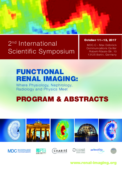
Functional Renal Imaging: Where Physiology, Nephrology, Radiology and Physics Meet
Dear colleagues and friends,
this symposium is designed to bring together scientists and clinicians from several disciplines, in a unique attempt to shape the future of renal functional imaging. It will provide an overview of cutting edge clinical and pre-clinical renal imaging techniques, and explore the clinical relevance of renal imaging, the future directions of renal functional MR, and the harmonization of these approaches with clinical applications.
We aim to provide a platform for fruitful engagement with colleagues and peers, and to foster the development of local, national and international collaborations to explore multi-disciplinary imaging approaches. The symposium is tailored to attract basic scientists, clinical scientists and clinicians from physiology, nephrology, radiology, internal medicine and related fields, as well as experts in imaging sciences and physics from all levels, ranging from students to advanced users and international experts. It is a symposium from the community for the community.
The scientific program comprises 13 sessions, covering a wide spectrum of renal physiology and pathologies, invasive quantitative approaches, optical imaging techniques, photoacoustic imaging and MR imaging. We are honored to present an array of outstanding international speakers including first class basic scientists, technology leaders and distinguished clinical experts. Focused sessions will provide deeper explanations into the most pressing imaging needs from the clinical perspective, and highlight the potential of renal imaging for the assessment of renal physiology, and the challenges en route to broader clinical applications.
Industry experts will present the current capabilities of functional MRI of the kidney, and outline the emergence of future renal MR imaging techniques. We aim to bridge disciplinary boundaries, and to engage the creativity and drive of the renal imaging community to solve the remaining challenges and overcome the barriers standing in the way of addressing unmet clinical objectives with renal functional imaging.
Poster sessions will run in parallel with the scientific program. We will also include multiple timeslots for poster power presentations, in order to bring to the attention of the audience a large number of poster presenters. Contributions of posters are strongly encouraged, and the best posters and poster power presentations, as judged by the audience, will receive an award.
We warmly invite you to join us for the 2nd International Symposium on Functional Renal Imaging in Berlin. Please save the date. A visit to Berlin is always worth it. Alongside the dynamic scientific landscape, Berlin has numerous historical landmarks, cultural highlights and sport events to offer.
We eagerly look forward to your participation. Be there – be Berlin!
- Previous meeting: 1st international meeting on renal MRI
- Next meeting: 3rd international meeting on renal MRI
- Abstracts and registration
-
-
Every registered participant is invited to submit one or several abstracts for poster presentations. Abstracts may be composed of a text with a maximum length of 650 words and up to 3 figures. The deadline for abstract submission is 27 August, 2017.
The abstract submission deadline has been extended until 4 September, 10:00 a.m. (Central European Summer Time).
At the symposium a board will be assigned to each poster presenter to display their poster. The poster should have A0 size in portrait format (poster display dimension 100 cm wide x 120 cm high).
During the symposium there will be several Power Poster sessions that will give a large number of poster presenters the opportunity to showcase their work in a 3-minute oral presentation. We are looking forward to your poster contributions, which will be all considered for the symposium’s poster price contest.
Please be aware that you have to register for the symposium before you will be able to submit an abstract.
If you have any questions, please directly contact us: renalsymp@mdc-berlin.de.
Venue
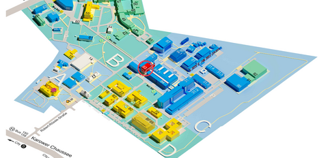
MDC.C Axon 1+2
Time
Catering
Lunch break
Coffee break
Social Event: Museum of Natural History
Program
Scientific Program
- Day 1
-
Wednesday 11 October 2017
-
- Download presentations (ZIP, 1.8 GB) (password required)
- Download selected presentations (password required)
Introduction
10:30
Welcome & objectives of meeting
10:40
The PARENCHIMA initiative: Aims and roadmap
Steven Sourbron – University of Leeds, UKRenal Physiology
10:55
Renal physiology: Urine formation and salt-water balance
Pontus Persson – Charité, Berlin, Germany11:15
Renal physiology: Renal oxygenation
Hans Joachim Schurek – Hannover Medical School, Germany11:35
Renal physiology: Regulation of intrarenal oxygenation
Roger Evans – Monash University, Melbourne, Australia11:55
Renal physiology: Regulation of renal perfusion
Erdmann Seeliger – Charité, Berlin, Germany12:15
Light lunch (posters)
Renal Diseases & Pathophysiology - Part I
13:00
Nephrological perspectives: Acute kidney injury (AKI)
Nicholas Selby – University of Nottingham, UK13:20
Nephrological perspectives: Diabetic nephropathy
Loreto Gesualdo – University of Bari, Italy13:40
Nephrological perspectives: Chronic kidney disease (CKD)
Alberto Ortiz – Autonomous University of Madrid, Spain14:00
The link between Cardiac and renal diseases
Ags Odudu – University of Manchester, UK14:20
Modelling renal pathophysiology: Promises and challenges of animal models
Andrea Fekete – Semmelweis University and Hungarian Academy of Sciences, Budapest, HungaryRenal MR Imaging Methods
14:40
Fibrosis and microstructure: T1 relaxation, apparent diffusion, diffusion tensor imaging
Neil Peter Jerome – Norwegian University of Science and Technology, Trondheim, Norway15:00
Oxygenation and blood volume: T2/T2* relaxation, BOLD, iron oxide enhancement
Andreas Pohlmann – Max Delbrück Center for Molecular Medicine, Berlin, Germany15:20
Perfusion and filtration: arterial spin labeling, dynamic contrast enhancement
Fabio Nery – UCL Great Ormond Street Institute of Child Health, London, UK15:40
Molecular imaging: Hyperpolarisation, magnetization transfer, CEST
Christoffer Laustsen – Aarhus University, Denmark16:00
Coffee break (posters)
(Minimally) Invasive Quantitative Measurements
16:30
The physiologists tool kit: Quantitative invasive probes
Kathleen Cantow – Charité, Berlin, Germany16:50
Renal optical methods: Near infrared spectroscopy
Dirk Grosenick – German Metrology Institute, Berlin, Germany17:10
Renal optical methods: Hyperspectral imaging
Wenke Markgraf – Dresden University of Technology, Dresden, Germany17:30
Renal optical methods: Phosphorimetric pO2 measurement
Philippe Guerci – University of Amsterdam, The Netherlands17:50
Renal optical methods: Optoacoustic renal imaging
Tim Devling – iThera Medical GmbH, Munich, Germany18:30
Management committee meeting of the COST action PARENCHIMA
Steven Sourbron – University of Leeds, UK - Day 2
-
Thursday 12 October 2017
-
- Download presentations (ZIP, 1.8 GB) (password required)
- Download selected presentations (password required)
Insights Into Dynamic Contrast-Enhanced MRI
09:00
What DCE MRI can(not) tell us about renal pathophysiology
Arvid Lundervold – University of Bergen, Norway09:20
Current challenges for using renal DCE MRI in the clinic
Nicolas Grenier – University of Bordeaux, FranceInsights Into Arterial Spin Labelling MRI
09:40
What ASL MRI can(not) tell us about renal pathophysiology
Charlotte Buchanan – University of Nottingham, UK10:00
Current challenges for using renal ASL MRI in the clinic
María Fernández Seara – Universidad de Navarra, Pamplona, Spain
10:20
Coffee break (posters)
Renal Diseases & Pathophysiology - Part II
11:00
A pharmaceutical industry’s perspective on how renal imaging can enrich clinical trials
Frank Czerwiec – Otsuka Pharmaceutical Development & Commercialization Inc., Washington D.C., USA11:20
What does the nephrologist expect from functional renal imaging
Kai-Uwe Eckardt – Charité, Berlin, Germany11:40
What does the pathophysiologist expect from functional renal imaging
Clive May – University of Melbourne, Australia12:00
What does the radiologist expect from functional renal imaging
Iosif Mendichovszky, Cambridge University Hospitals, UK
12:20
What does an expert radiographer expect from renal imaging
Eli Eikefjord – University of Bergen, Norway12:40
Lunch break (posters)
Insights Into Oxygenation MR Imaging
14:10
What oxygen sensitive MRI can(not) tell us about renal pathophysiology
Pottumarthi Vara Prasad – NorthShore University HealthSystem, Evanston, USA14:30
Current challenges for using renal BOLD MRI in the clinic
Lilach Lerman – Mayo Clinic, Rochester, USARenal Power 1 - Clinical Research
(13 presentations of 3 min each)
14:50 Acute pyelonephritis in children: Diagnostics and comparison of two methods – static renal scintigraphy and MR imaging
Alice Bosáková – University Hospital Ostrava-Poruba, Czech RepublicRole of image and clinical-based biomarkers in renal transplant assessment
Mohamed Abou El-Ghar – Urology & Nephrology Center, Mansoura University, EgyptThe effect of enhancing spatial resolution in non-contrast enhanced renal MR angiography
Anne Dorte Blankholm – Aarhus University Hospital, DenmarkReference method for measurement of total renal function - estimated versus measured GFR
Martin Šámal – First Faculty of Medicine, Charles University & General University Hospital, Prague, Czech RepublicThe cortico-medullary ADC difference reduces inter-system variability in renal diffusion-weighted imaging
Iris Friedli – Geneva University Hospitals, University of Geneva, Faculty of Medicine, SwitzerlandFeasibility study of diffusion tensor imaging of the kidneys in freely breathing infants with pyelonephritis
Yvonne Simrén – Institute of Clinical Sciences at Sahlgrenska Academy, University of Gothenburg, SwedenValidation of single-kidney glomerular filtration rate measurement with dynamic contrast-enhanced MRI
Susmita Basak – Division of Biomedical Imaging, LICAMM, University of Leeds, UKRepeatibility of renal BOLD MRI
Anneloes de Boer – University Medical Center Utrecht, The NetherlandsFeasibility of renal ASL in a pediatric cohort with impaired renal function
Fabio Nery – UCL Great Ormond Street Institute of Child Health, London, UKKidney fibrosis assessment: T1rho and DCE permeability study
Dmitry Kupriyanov – Philips Healthcare, Moscow, RussiaA pilot, multi-vendor comparison of multi-echo gradient-echo acquisitions for BOLD imaging in the kidney
Pim Pullens – University Hospital Brussels, BelgiumDiffusion weighted MR imaging to investigate response to therapy: A case report
Anna Caroli – IRCCS - Istituto di Ricerche Farmacologiche Mario Negri, Bergamo, ItalyVariability reduction in renal diffusion-weighted MR imaging with motion compensation
Iris Friedli – Geneva University Hospitals, University of Geneva, SwitzerlandRenal Power 2 - Experimental Research
(7 presentations of 3 min each)
15:35
The renal effect of anesthesia on the functional and metabolic phenotype in rats
Haiyun Qi – MR Research Centre, Department of Clinical Medicine, Aarhus University, DenmarkSimultaneous assessment of kidney perfusion and pH in an acute kidney injury murine model exploiting a dynamic CEST-MRI approach
Dario Longo – National Research Council of Italy, Rome, ItalyAssessment of metabolism in early renal ischemia/reperfusion injury using hyperpolarized 13C-pyruvate
Per Mose Nielsen – Department of Clinical Medicine - Biomedical Radio Isotope Techniques, Aarhus, DenmarkMeasuring renal oxygenation in a mouse model of volume-dependent hypertension using BOLD MRI
Dexter Lee – Howard University, Washington D.C., USAQuadrature birdcage RF coil for renal sodium (23Na) imaging in rodents at 9.4 T: Initial results
Laura Böhmert – Max Delbrück Center for Molecular Medicine, Berlin, GermanyNoninvasive evaluation of renal pH homeostasis after ischemia reperfusion injury by CEST-MRI pH mapping
Dario Longo – National Research Council of Italy, Rome, ItalyDiffusion-weighted split-echo RARE imaging free of geometric distortion for renal MRI at ultrahigh fields
João dos Santos Periquito – Max Delbrück Center for Molecular Medicine, Berlin, Germany16:00
Poster session (coffee break) Insights Into Diffusion MR Imaging
16:50
What diffusion MRI can(not) tell us about renal pathophysiology
Jean-Paul Vallee – University of Geneva, Switzerland17:10
Current challenges for using renal diffusion MRI in the clinic
Alexandra Ljimani – University of Düsseldorf, GermanySocial Event
19:00
Social Event
- Day 3
-
Friday 13 October 2017
-
- Download presentations (ZIP, 1.8 GB) (password required)
- Download selected presentations (password required)
Insights Into Emerging MR Technologies
09:00
Susceptibility MR: Better and bolder than BOLD
Chunlei Liu – University of California, Berkeley, USA09:20
Sodium MR is worth its salt!
Stefan Zbyn – University of Oulu, FinlandRenal Power 3 - Multiparametric Studies
(9 presentations of 3 min each)
09:40
Assessment of renal stiffness in IgA nephropathy using multifrequency MRE compared to DWI and BOLD
Stephan Marticorena Garcia – Charité - Universitätsmedizin Berlin, GermanyMultiparametric kidney MR imaging to identify novel markers of disease progression in autosomal dominant polycystic kidney disease
Albert Ong – University of Sheffield, UKMolecular DCE MRI with high and low temporal resolution, using bimodal AGulX contrast agents and multiparametric MRI in a murine UUO model
Gregory Ramniceanu – Chimie ParisTech, Unité de Technologies Chimiques et Biologiques pour la Santé, FranceMultiparametric MRI of renal transplant: Preliminary comparison of advanced MRI parameters in patients with functional and fibrotic renal allografts
Octavia Bane – Icahn School of Medicine at Mount Sinai Hospital, New York, USAMR imaging to assess the pathophysiology of acute kidney injury
Huda Mahmoud – University of Nottingham, UKHyperpolarized [13C,15N2]urea: A novel renal O2 saturation biomarker in acute kidney injury?
Christian Mariager – Department of Clinical Medicine - Biomedical Radio Isotope Techniques, Aarhus, DenmarkNon invasive MRI of renal physiology
Per Eckerbom – Institute for Surgical Sciences, Uppsala University, SwedenMultiparametric assessment of chronic kidney disease
Charlotte Buchanan – University of Nottingham, UKEvaluation of fibrosis models using 1H T1 mapping and slow component T2 23Na
Per Mose Nielsen – Department of Clinical Medicine - Biomedical Radio Isotope Techniques, Aarhus, DenmarkRenal Power 4 - Segmentation & Modelling
(5 presentations of 3 min each)
10:10
Novel strategy for contrast-free MR angiography of arteriovenous fistulae for hemodialysis
Andrea Remuzzi – University of Bergamo, Bergamo ItalySemi-automated kidney delineation on BOLD images using k-means clustering of R2* signal decay
Anneloes de Boer – University Medical Center Utrecht, The NetherlandsFully automatic kidney segmentation in abdominal MR imaging using random forests (RFs)
Marc Fischer – University of Tübingen, GermanyThe influence of hydration status of kidney volume and cyst measurements
Jens Dam Jensen – Department of Clinical Medicine - Biomedical Radio Isotope Techniques, Aarhus, DenmarkFast semi-supervised segmentation of the kidneys in DCE-MRI using convolutional neural networks and transfer learning
Alexander Lundervold – Western Norway University of Applied Sciences, Bergen, Norway10:30 Poster session (coffee break) The Industry's Perspective
11:10
Renal imaging in pharmaceutical research: Investigating glomerular pathophysiology using micro-CT
Luke Xie – Genentech, San Francisco, USA11:30
State-of-the-art and future directions in preclinical abdominal MRI
Claudia Oerther – Bruker Biospin GmbH, Ettlingen, Germany11:50
Clinical MRI of the kidneys – Today and tomorrow
Hans Peeters – Philips Healthcare, Eindhoven, The NetherlandsLooking Beyond The Horizon
12:10
Improving diagnostic accuracy and treatment of renal diseases with computational models
Anna Caroli – IRCCS - Istituto di Ricerche Farmacologiche Mario Negri, Ranica - Bergamo, Italy12:30
Predictive analytics and machine learning for advancing renal diagnostics and therapies
Kolja Bailly – Solandeo GmbH, Berlin, Germany12:50
Practical challenges of multi-center studies and clinical renal imaging trials
Paul Hockings – Antaros Medical, Gothenburg, Sweden13:10
Adjourn (symposium)
13:20
Lunch break
Working Group Meetings organized by COST action PARENCHIMA
Plenary session14:00
Introduction: Organization of the WG meeting
Steven Sourbron – University of Leeds, UK14:05
Working Group 1 pitch: objectives, progress, task forces, questions
Reproducibility and standardization
Christoffer Laustsen – Aarhus University, Denmark14:20
Working Group 2 pitch: objectives, progress, task forces, questions
Research and development toolbox (open-access toolbox with software & data)
Frank G. Zöllner – Heidelberg University, Germany14:35
Working Group 3 pitch: objectives, progress, task forces, questions
Multi-centre clinical trial
Anna Caroli – IRCCS – Istituto di Ricerche Farmacologiche Mario Negri, Ranica – Bergamo, Italy14:50
Working Group 4 pitch: objectives, progress, task forces, questions
Training programs
Thoralf Niendorf – Max Delbrück Center for Molecular Medicine, Berlin, Germany15:05
Working Group 5 pitch: objectives, progress, task forces, questions
Dissemination (incl. PARENCHIMA Action website, intl. meetings)
Marcos Wolf – Center for Medical Physics and Biomedical Engineering, Vienna, AustriaWorking Group Meetings organized by COST action PARENCHIMA
Breakout sessions15:30
Working Group 1: Improving the reproducibility and standardization of renal MRI
Lead: Christoffer Laustsen, Aarhus University, Denmark15:30
Working Group 2: Development of a renal MRI open-access toolbox with software & data
Lead: Frank Zöllner, Heidelberg University, Germany15:30
Working Group 3: Multi-centre clinical trial
Lead: Anna Caroli, IRCCS – Istituto di Ricerche Farmacologiche Mario Negri, Ranica – Bergamo, Italy15:30
Working Group 4: Development of a training program on renal MRI for basic scientists and clinical users
Lead: Thoralf Niendorf, Max Delbrück Center for Molecular Medicine, Berlin, Germany16:30
Reporting: Roles, planning, milestones, STSMs
17:15
Internal sum up per Working Group
(task forces present their report; reports will be collated into a summary for newsletter/website)17:30
Adjourn
- Download presentations (ZIP, 1.8 GB) (password required)
- Download selected presentations (password required)
Fees
Regular fee
- Early (until 28.07.17): 300 €
- Late (until 29.09.17): 400 €
- On-site: 450 €
Student fee (incl. PhD students)
- Early (until 28.07.17): 150 €
- Late (until 29.09.17): 190 €
- On-site: 200 €
Day ticket
- Per day: 200 €
Organizers
Andreas Pohlmann (MDC)
Erdmann Seeliger (Charité)
Dirk Grosenick (PTB)
Sonia Waiczies (MDC)
Kathleen Cantow (Charité)
Pontus Persson (Charité)
Thoralf Niendorf (MDC)
Co-organizers
This symposium is co-organized by the European Cooperation in Science and Technology (COST) action 'MRI Biomarkers for Chronic Kidney Disease' (PARENCHIMA, CA16103).
Contact
Rosita Knispel
phone: +49 30 9406 4505
Lien-Georgina Dettmann
phone: +49 30 9406 2719


