
Treier Lab
Genetik metabolischer und reproduktiver Störungen
Tissue Clearing and Light Sheet Microscopy
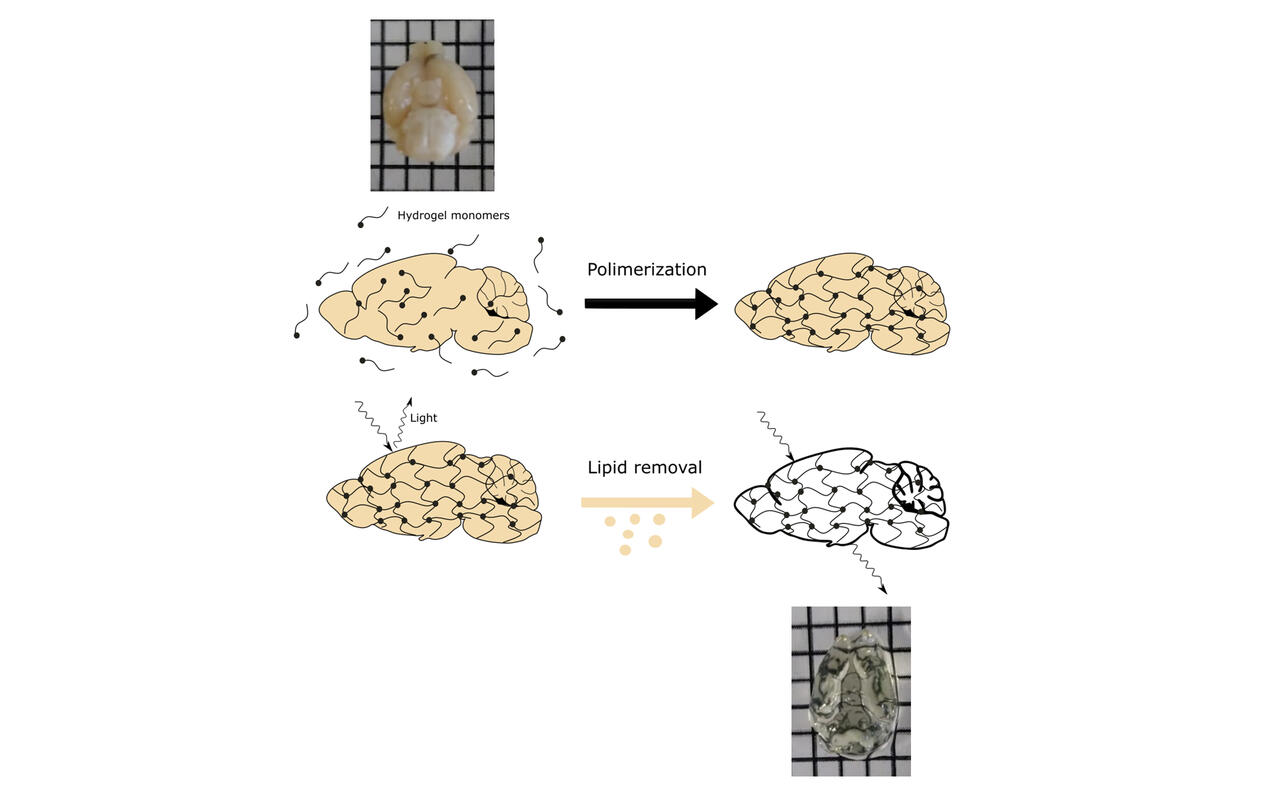
CLARITY
CLARITY stands for Clear, Lipid-exchanged, Anatomically Rigid, Imaging compatible, Tissue hYdrogel and is a technique developed by the Deisseroth lab in Stanford, California, and published in May 2013 in Nature.
With this state-of-the-art method in neuroscience it is possible to turn a brain transparent using the detergent SDS, which strips away lipids that normally block the passage of light. First, the brain is infused with acrylamide which binds proteins, nucleic acids and other biomolecules. After heating the acrylamide polymerizes and forms a tissue-wide mesh that secures the molecules. By using this technique it is possible to visualize and trace, quantify and map the axonal projections of the histaminergic neurons in an intact system in whole fluorochrome-expressing brains.
Light Sheet Microscopy (LSF)
Light Sheet Microscopy has become the method of choice when imaging cleared tissue such as the murine brain at cellular level. In contrast to other imaging systems like confocal microscopy or 2-photon microscopy, during LSM the sample is illuminated in form of a thin sheet of light perpendicular to the detection objective. The emitted fluorescent light is collected by the objective and projected to fast capturing image sensors like CCD or sCMOS cameras. The excitation of only a thin tissue slice combined with high speed acquisition minimizes bleaching while providing an intermediate to high resolution.

