Darth Vader’s helmet and other adventures at the border of physics, medicine, and biology
Suppose you have a free afternoon to spend discussing topics like interdisciplinarity, new technology and translational research, with physicists, biologists, doctors, patients, computer programmers, and possibly a guy who drives a crane. And while you’re at it, why not fool around with a 32-ton magnet?
All that fun and more can be had by hanging out with Thoralf Niendorf’s group at the Berlin Ultrahigh Field Facility (B.U.F.F.), with its impressive MRI machines. If you don’t know where that is, just walk around campus with a compass, or some loose change in your pocket, and follow it to the lovely white cube across from the ERC, where the bus enters campus. There you’ll find not only impressive equipment – including one of the largest MRI machines in Germany – but also a creative team that is turning a rather blunt instrument into a more finely honed tool for biomedical research.
MRI is considered one of the most important technologies ever developed for medical diagnosis. The campus facility was set up as a vision of former Scientific Directors Walter Birchmeier and Walter Rosenthal, but its methods have yet to be truly integrated into most mainstream molecular biology research. For an imaging technology, that’s not unusual – remember the on-again, off-again reputation of light microscopy before the great revolution in fluorescent proteins? They ushered in the age of confocal microscopy, and now digital sheet light microscopy, and techniques such as FRET, FRAP, FLIM, and exquisite new methods to study the biophysical properties of molecules and cells in vivo.
Similarly, innovations in MRI may soon lead to new uses for the methods in the laboratory and clinic. For many, the main barrier has been spatio-temporal resolution. While “classical” MRI has permitted an in vivo view of tissues deep within the brain and other internal organs, it hasn’t reached a resolution capable of imaging cells or, of course, molecules. But Thoralf’s group is sharpening the picture thanks to the arrival of ultrahigh field MRI (UHF-MRI), which produces stronger magnetic fields, and generates higher radio frequencies that can be precisely focused using custom-made antenna arrays and can be tuned to visualize sodium ions as well as hydrogen.
Thoralf and his group are internationally recognized for a number of these innovations in UHF-MRI, some of which have already been implemented as clinical tools in medical diagnoses. The group has embarked on several interesting multi-disciplinary projects that will make it possible to capture new types of information – at a higher level of detail – from living tissues. Their work has led to new uses for the technology in research, phenotyping, diagnosis, and potentially new types of treatments. While the MRI building will remain on the fringe of the Buch campus, their work will not – if the group and their colleagues at the MDC can bridge some interdisciplinary gaps.
* * * * *
On a sunny Tuesday afternoon Thoralf invites me to arm wrestle with the 7-Tesla MRI machine, whose 32-ton magnet emits a field that would wipe your credit cards, stop your watch, and reach for anything metallic you forgot to remove from your pockets. I imagine all the lost keys on campus slowly creeping toward the building.
“What about the fillings in my teeth?” I say. They should be all right, he says.
He turns on the machine, which emits a series of loud, regular clicks familiar to anyone who has ever taken a ride into the tube of an MRI scanner. Now come a series of physical experiments involving a hoop, a bar, and a plate the size of a large book, all made of metal. Thoralf places the plate in front of the machine, where it levitates and tilts off in one direction; he challenges me to tip it over. I place my palm against it and push, hard, and can only budge it a bit. In contests of brute force, the laws of physics always win; 7-Tesla retains its title as campus arm-wrestling champ.
MRI, I consider, might be the reason Thor’s hammer can’t be lifted, or Arthur’s sword pulled from the stone.
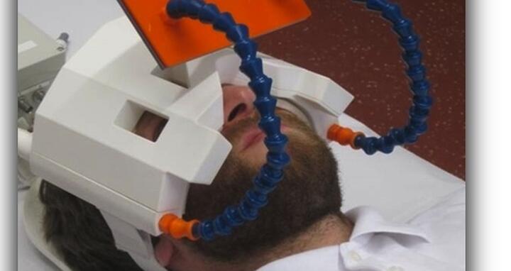
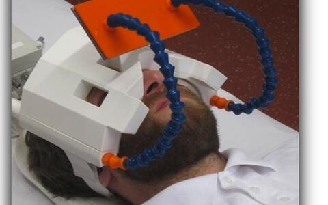
After that bit of fun, Thoralf rummages around in a box near the machine and pulls out a strange piece of gear, white and head-shaped, with antennae-like extensions. “We call it Darth Vader’s mask,” he says. “We built it to solve a problem raised by an ophthalmologist from the University of Rostock and a radiologist from the University of Greifswald.”
The question involved capturing detailed images of the eye, and this required solving two challenges. The first was, once again, resolution: the radio frequencies generated in classical MRI instruments are long waves, which lead to large “pixels” in images. Sharpening the focus requires higher frequencies with shorter wavelengths, which have to be focused using multiple radio transmitters. Hence the six radio antennae, which are positioned in front of the eyes during a scan.
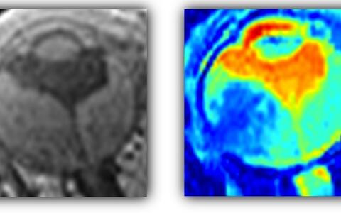
The second challenge involved what the physician was hoping to see. Small ocular masses within the fluid of the eye usually indicate the presence of a small tumor or some other irregularity. They are tiny and virtually indistinguishable from the healthy matter of the eye, using traditional MRI techniques, because the machine images the fluid content of tissues – the water they contain – and can’t pick out the masses from the background.
Thoralf and Katharina Paul, who is a PhD student in his group, found a solution by focusing on a biophysical characteristic of fluids: Brownian motion. At the atomic scale, matter is always in motion. The atoms and molecules in liquids are always performing a frenetic dance, as a consequence of random forces acting on them. When linked to each other within solid structures, this motion is more restricted. The group found a way to resolve these differences in images from the UHFMRI machine, turning it into the first remote detector for subtle tumors of the eye.
Within two weeks after the completion of the project, Thoralf got a call from the Charité – from physicians wanting to use the technology in the planning of treatments for patients using the proton beam at the Helmholtz Center in Berlin. Thoralf jumped onto this opportunity right away because of the outstanding framework for clinical collaborations in Berlin. “This provides unique opportunities for translating our technologies into clinical applications, in fair and mutual collaborations,” Thoralf says.
* * * * *
The group has faced some difficulties in terms of campus integration because many scientists find it difficult to grasp the creative and collaborative efforts needed to solve particular problems in MRI.
“MRI cannot be used as a microscope,” Thoralf says. “You don’t just come with a sample – in this case a living subject – and stick it in the machine, and out pop the images you’re after in realtime.”
Instead, he says, scientists need to think of the technology the way they would regard small-molecule screening, where weeks or months of work are often required to perfect an assay before carrying out a genome-wide screen. With MRI the challenges involve the physiology of the tissue to be studied: its size and location in the body, and the type of biological processes that scientists wish to observe.
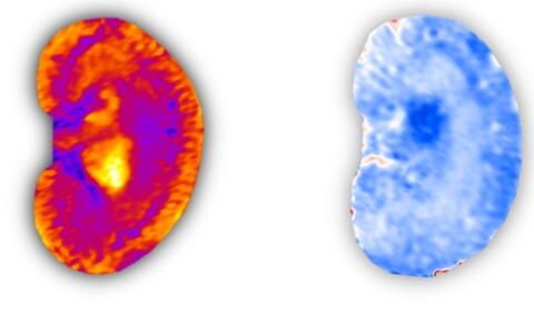
Collaborators from the Charité hoping to study the kidney, for example, wanted to observe blood flow and the oxygenation of tissues. Thoralf and Andreas Pohlmann from his team found a solution which also allows scientists to distinguish different types of fat that are indicative of various pathological conditions. These activities have culminated in the team’s contributions to the DFG funded research group on the haemodynamics of acute kidney injury, which are now being taken to the next level by an SFB proposal initiative. In general, he says, 7-Tesla MRI is an excellent method to study the circulatory system, permitting a detailed view of the growth and structure of blood vessels during the development of healthy and cancerous tissues.
One of the major goals has been to capture images of the working heart, to study its functions in living organisms during the progress of various types of cardiovascular diseases. This has required, for example, the development of sophisticated antenna designs – but also tuning the instrument to track sodium ions, rather than hydrogen.
“Changes of sodium concentrations have been linked to disturbances in cellular homeostasis and a loss of tissue viability,” Thoralf says. “But the sodium concentration in tissues is about 2000 times lower than that of hydrogen, which causes problems when you’re trying to pick out a signal against the background noise.”
Sodium concentrations had been observed using various combinations of radio frequency (RF) antennae and arrays to focus signals on specific targets in the body. But these arrangements had provided only very focused views of particular regions of organs. What was needed was an array that could scan large areas – such as the heart, or entire body. The solution was found in a collaboration with Armin Nagel from the DKFZ Heidelberg and Friedrich Wetterling from the Trinity College in Dublin within the framework of the ICEMED (imaging and curing environmental and metabolic diseases), a Helmholtz alliance. They built a new system of RF detector coils and a resonator system that could excite sodium ions in a highly uniform way, across the entire body.
The collaboration produced the first images of the entire, beating heart that clearly delineated healthy and diseased tissues – at a temporal resolution of 30 microseconds. Developing the method further, in a way that can easily be implemented in clinics, would provide a valuable new tool for the diagnosis of heart disease. That will require more work – which Friedrich aims to carry out within Thoralf’s group. He has just submitted an application for a Marie-Curie grant in hopes of funding the project.
These efforts have improved spatial resolution for visualizing the torso and organs such as the lungs and heart by a factor of ten. The same basic method can be used to visualize microstrokes, very local bleeding within the brain.
A project that critics originally laughed at – believing that the physics problems could not be solved – has made the group leaders in the use of 7-Tesla machines for cardiac imaging, thanks to a collaboration with Jeanette Schulz-Menger and other cardiologists from the ECRC.
* * * * *
The list of innovations from the lab goes on and on. One of Thoralf’s early projects, before his arrival at the MDC, was to use MRI to diagnose multiple sclerosis.
“There was no definitive method to distinguish MS from a large range of other neurological conditions,” Thoralf says. “But a link had been established between the density of blood vessels and the severity of MS. That was something we could visualize using MRI, and it provided a clear view of lesions associated with the progression of the disease. So this gave us the first method to make definitive diagnoses in this terrible, degenerative condition thanks to the clinical collaboration with Friedemann Paul and his team from the excellence cluster NeuroCure.”
Another project has been to study fluorine – which is interesting because the body doesn’t produce it. Observing the uptake of fluorine can give insights into a range of, cellular, pharmacological, metabolic and other processes. MRI of fluorinated cells provides a unique means to shed light on neuro-immunological diseases, which has been demonstrated by Sonia Waiczies, who is a senior scientist in Thoralf’s group. Once again, detecting the substance lies at the very edge of MRI capacities. Thoralf and Sonia found a solution by designing a new, helium-filled cryo-coil – and convinced a manufacturer to develop it for clinical use.
The group’s PhD students are pursuing a number of interesting projects. Henning Reimann has just designed a “cradle” that maintains normal physiological temperatures in mice during an MRI experiment studying brain activity. That’s particularly important, he says, in studies of animal pain that use heat as a stimulus.
“Mice are anesthetized during MRI, which changes some of the parameters of their base body temperature,” Henning says. “Their bodies adapt to the environment, which might be a problem if you’re using heat to trigger a response – the background temperature of a tissue might influence the way it reacts to the stimulus.”
The new cradle permits measuring – and precisely maintaining – temperatures throughout the animal body as such studies are carried out. At the very least, it’s an important control for experiments studying an animal’s response to pain.
And a new project by Lukas Winter, also the subject of a grant application, aims to turn MRI into a method of introducing precisely focused heat into internal tissues in living animals. This would open the door on entirely new types of studies of thermoregulation, using temperature to phenotype tissues and pathological processes, and studying the effects of temperature on metabolism and other processes. It might even offer a new type of therapy, based on heating up diseased tissues rather than bombarding them with radioactive ions. Thoralf and Lukas are driving this from blue sky explorations into the physics behind this phenomenon into clinical reality in collaboration with Peter Wust from the department of radiation oncology of the Charité.
* * * * *
“It is no secret that the future of MRI will not end at 7.0 T,” Thoralf says. “The field is moving in a direction involving even larger magnets and more sophisticated arrangements of antenna arrays.
In his wildest dreams, Thoralf says, he would be keen to attract the world’s first 20 Tesla human MRI scanner to the MDC campus. While for the moment this merely a vision, it would permit scientists to carry out MRI of sodium with a sub-millimeter spatial resolution in 5-10 min scan time, and offers the potential for probing single cells in vivo.
In view of this goal, recent pioneering simulations from Thoralf and Lukas Winter have demonstrated the potential of human in vivo MRI at 14 Tesla (600 MHz) and 23.5 Tesla (1 GHz), the world’s first reports of this kind. This exclusive expertise led to an invitation to Thoralf to join his American colleagues for a strategic workshop entitled “Ultrahigh Field NMR and MRI: Science at the Crossroads,” to be held in November at the NIH, in Bethesda, Maryland. The purpose of the meeting is to establish, as a community, a roadmap for the development of ultrahigh magnetic field technologies, and raise awareness among funding agencies with the aim of procuring of multiple instruments in key shared instrumentation centers across the United States.
“We need similar enthusiasm and momentum among the European imaging community; I am convinced of the inevitability and power of higher magnetic field strengths. That’s why I launched an annual symposium on ‘Ultrahigh Field Magnetic Resonance: Clinical Needs, Research Promises and Technical Solutions,’ which is open to all who are interested.”
I leave the facility with the impression that this is an intensely creative, innovative group on campus, actively pushing an imaging technology beyond its current limits to cope with new scientific questions. The projects are complex, time-consuming, and in some cases expensive. But Thoralf and his colleagues are well on their way to turning MRI into a mainstream tool for molecular biologists who aim to study the effects of molecular processes at scales ranging from cells to tissues, in living organisms. All that’s needed are groups willing to invest time and energy into exploring this higher dimension of health and disease.
Featured Image: "Thor(alf)'s hammer:" Thoralf Niendorf preparing for a wrestling match with the 7-Tesla MRI scanner. (Warning: DO NOT try this with your own MRI machine because it constitutes a severe safety hazard! Here the magnet was off during installation; after installation, the magnet is ALWAYS on.) Photo: Sabrina Klix, MDC


