Heart muscle cells merge when the heart grows and repairs itself
Cells fuse for various reasons. We know that skeletal muscle fibers develop from merged progenitor muscle cells. Cell fusion also seems to play a role in disease processes such as cancer. Being able to reprogram differentiated cells by fusion to encourage growth offers a new way forward for treating many diseases. Yet the mechanisms behind fusion are not well understood and the significance of this process is unclear. In addition, models and tools capable of observing fusion in living organisms under normal conditions have so far been lacking.
Heart muscle cells fuse temporarily
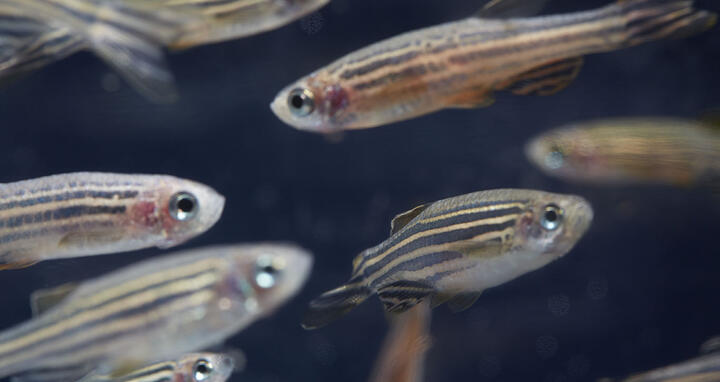
Dr. Suphansa Sawamiphak, a researcher at the German Center for Cardiovascular Research (DZHK), and her colleagues at the Max Delbrück Center for Molecular Medicine in the Helmholtz Association (MDC) in Berlin and at the Max Planck Institute for Heart and Lung Research in Bad Nauheim have now succeeded in genetically altering zebrafish larvae so that they can reliably detect cell fusions in a living organism. They have dubbed their system “Fluorescence Activation After Transgene Coupling,” or FATC for short. A fluorescent reporter gene is read only when the cells are fused. These cells then light up when viewed under a fluorescence microscope. Researchers have used this to observe heart muscle cells fusing in fish for the first time. These are transient fusions: the cells glow only as long as they are connected to each other. When they divide again, the light signal is lost.
“What’s so special about this is that we can use it to visualize system fusions throughout the body,” says Sawamiphak. “Until now, you could only observe cell fusions by transplanting marked cells such as bone marrow cells and then monitoring to see whether the cells fuse in certain tissues. The tissue and cell type were predetermined, so unknown fusion processes remained hidden.”
In the development phase, an unexpected discovery
Wounded zebrafish are able to completely regenerate themselves. The Berlin researchers observed that heart muscle cells fuse together during this phase. They found evidence for this process during fish larvae development. “We expected fusions during regeneration of a wounded heart. But it was astonishing to see it happening during development,” says Sawamiphak.
Cell growth goes hand in hand with fusion
Further experiments showed that the proportion of fusion cells dropped by more than half in the hearts of the adult fish compared to the larvae hearts. “The healthy adult heart muscle also has little cell growth,” says Sawamiphak. “So we examined whether the transient cell fusions were linked to cell division activity.” The results show that fused heart muscle cells divide very actively during cardiac development. The researchers also observed this relationship between cell growth and fusion in the adult fish heart when it regenerated itself. After an injury, the rate of cell division in a zebrafish heart generally increases greatly. And in genetically modified zebrafish the fluorescent—and thus fused—cells accounted for a significant portion of these actively dividing cells in the heart. Sawamiphak suspects that the heart muscle cells fuse temporarily in preparation for cell division.
Understanding regeneration and leveraging this knowledge
Next, Sawamiphak and her team want to find out exactly how fusion and cell division are related to each other. They also plan to conduct an analysis of the cell fusion mechanisms. Looking ahead, the researcher says: “Our findings show that there are fundamental mechanisms in the zebrafish. We hope this will help us better understand how the zebrafish heart regenerates, and can then use this knowledge to find new ways of treating damaged mammalian hearts.”
Further information
Sawamiphak S. et al. Transient Cardiomyocyte Fusion Regulates Cardiac Development in Zebrafish. Nature Communications 8, 1525, (2017). DOI: 10.1038/s41467-017-01555-8


