Looking inside the heart
Thoralf Niendorf
Magnetic resonance imaging (MRI) allows scientists to see what happens inside the body by watching organs at work. The technology uses a very strong magnetic field and radio waves to produce a series of “slices” of the body that depict different types of tissue. The images provide information about the anatomy and function of organs and vessels. MRI exams help to diagnose disease by visualizing damage in, for instance, the heart or the brain.
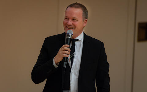
When talking about MRI scanners with a magnetic field strength of 7 Tesla or more, researchers use the term ultrahigh field magnetic resonance (UHF MR) imaging. The resolution of the images is so sharp that details measuring as little as half a millimeter are visible. “A 7-Tesla MRI scanner delivers more horsepower than conventional clinical systems. This increase in sensitivity can – like an HD television – be translated into sharper images, which help physicians identify diseases better,” says Professor Thoralf Niendorf of the MDC. This year marked the tenth year that Niendorf and the team at the Berlin Ultrahigh Field Facility (B.U.F.F.) have held the UHF MR symposium at the MDC. The other organizers were the Weizmann Institute of Science in Rehovot (Israel), Charité – Universitätsmedizin Berlin, and the Physikalisch-Technische Bundesanstalt in Berlin. The symposium was supported by the German Research Foundation (DFG) and Stiftung Charité.
MRI pioneers get together
Kamil Ugurbil
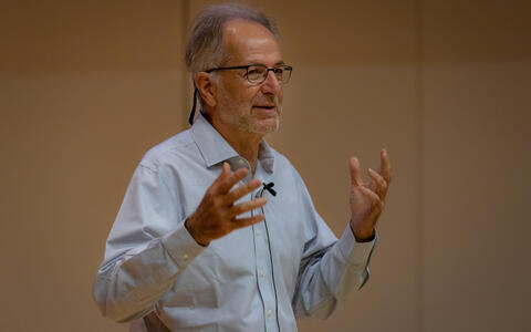
At the beginning of the event, Professor Kamil Ugurbil showed images of the prostate, hips, torso, and kidneys. He explained that no MRI scanner had ever before produced such high-resolution images of human organs. Ugurbil’s work at the University of Minnesota currently involves using the world’s most powerful MRI scanner for humans, which has a magnetic field strength of 10.5 Tesla. An extraordinary feature of brain function, said Ugurbil, is the encoding of information at multiple spatial and temporal scales, going from cellular level processes to coordinated activity over billions of neurons spanning large parts of it, if not the entirety of the brain. “Our overarching goals has been to push the limits of MR imaging of anatomy, function and connectivity to cover as much as possible of this vast operational scale, aiming for whole brain coverage necessary to study large scale networks but with sufficient resolution to access at the same time mesoscale and sub-mesoscale organizations that perform elementary computations”, said Ugurbil. “Very high magnetic fields, starting with 7 Tesla and now 10.5 Tesla are indispensable tools in reaching this aim. At the same time, these imaging capabilities are sure to improve our studies of other organ systems in the human body.”
We specifically want to give young scientists the opportunity to present their work and talk to us about it.
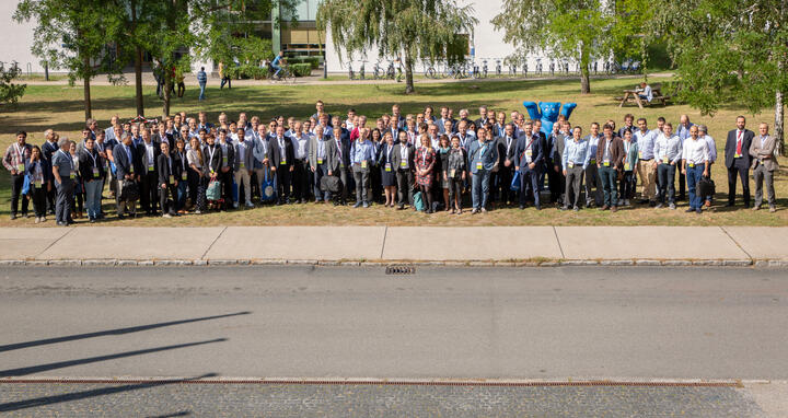
The symposium on September 6, 2019, brought researchers together to report on current developments and discuss the future of UHF MR. Seventeen speakers made up the scientific program, and 23 researchers exhibited posters depicting their current research.
Professor Armin Nagel from Friedrich-Alexander Universität ErIangen-Nürnberg gave attendees insights into heart muscle cells. A research collaboration with the Niendorf Lab at the MDC allowed Nagel to capture a potassium signal in a living human heart for the very first time. This was not possible with earlier generations of MRI scanners or other imaging modalities.
Professor Armin Nagel from Friedrich-Alexander Universität ErIangen-Nürnberg gave attendees insights into heart muscle cells. A research collaboration with the Niendorf Lab at the MDC allowed Nagel to capture a potassium signal in a living human heart for the very first time. This was not possible with earlier generations of MRI scanners or other imaging modalities.
Former MDC student wins poster competition
During the symposium attendees were asked to choose the best poster on display. “We specifically want to give young scientists the opportunity to present their work and talk to us about it,” said Niendorf. The most votes went to Eva Peper for her poster about measuring blood flow in the heart, the aorta, and the carotid artery. She developed a new technique for performing MR scans faster. This was the second time that Peper had attended the UHF MR symposium. She wrote her master’s thesis in the Niendorf Lab and is currently working as a doctoral candidate at Amsterdam UMC in the Netherlands.
Text: Christina Anders


