The world of protein crystals – How scientists of the MDC make the molecular world visible
The small crystals are shimmering under the microscope. As beautiful as they look, they won’t disclose their secrets on first sight. Technically they consist of millions of little protein molecules that are so tiny, even the highest resolution light microscopes fail to make them visible. But in order to understand how proteins work in the human body and communicate with each other, scientists have to find out how they look like. That’s where X-ray crystallography comes into play. It’s one of the most important methods for determining the molecular structure of proteins.
We talked with Manuel Hessenberger, Arthur Alves de Melo and Stephen Marino of the AG Daumke to find out more about their research and crystal structure analysis. At the Long Night of Sciences on 13 June 2015, visitors can take part in lab tours where the scientists will explain how molecular machines in the cell work and which mechanisms are behind the body’s immune defence against viruses.
In the AG Daumke X-ray crystallography has been used to study the structure of therapeutic cancer antibodies. Stephen Marino has been part of a project to develop and optimise these new antibodies to recognise and kill tumour cells. Now they are being evaluated by pharma companies for potential development into a treatment for a fatal blood cancer (multiple myeloma).
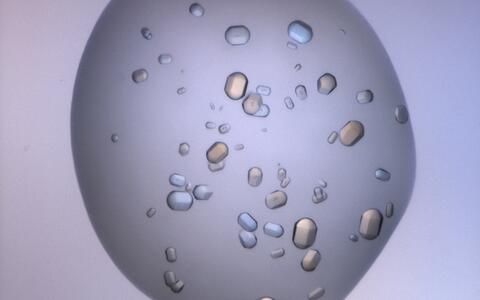
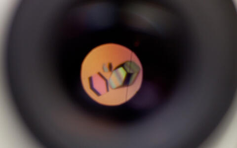
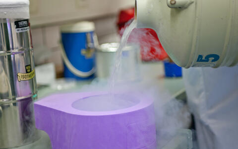
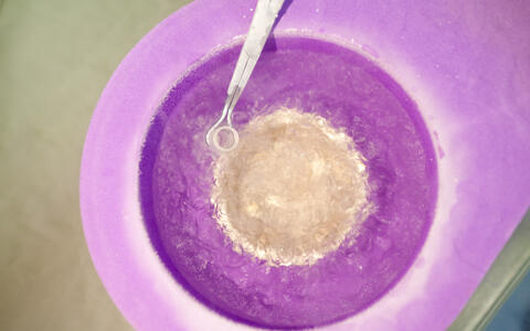
In our gallery below, Manuel Hessenberger, Arthur Alves de Melo and Stephen Marino give us a step by step explanation of the process of obtaining a 3D model of a protein molecule by X-ray crystallography.
Making the invisible visible
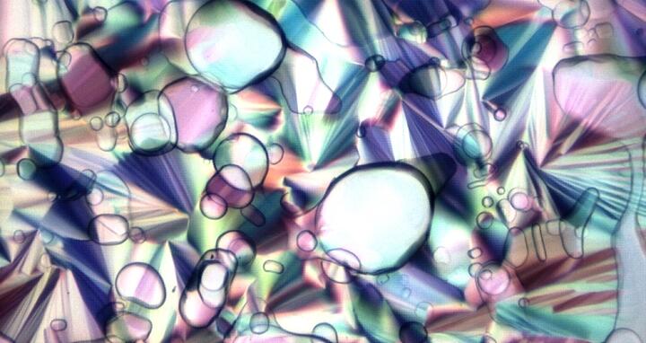
Featured Image: Light is reflected from a small droplet - the 200 billionth part of a liter – of hydrated lipid molecules, where special types of proteins can crystallize. Photo: © Dr. Stephen Marino, MDC

