
The Best Scientific Pictures
Science is beautiful: we take this literally

This confocal image shows epicardial adipose tissue (eAT), the fat depot on the surface of the myocardium, in an adult zebrafish heart. Zebrafish eAT closely resembles metabolically active fat found in humans. Model organisms, such as zebrafish, give insights into adipocyte biology and its implications for human health. Staining with BODIPY for lipids/adipocytes in yellow, DAPI for nuclei in blue, a transgenic line kdrl: HRAS-mCherry for the coronary vascular system in magenta, and acetylated tubulin for innervation in cyan.

A vivid image of cancer cells invading healthy tissue, like an inferno. Such detailed visualization by immunofluorescence microscopy can help unravel how cancer cells interact with the surrounding microenvironment and contribute to treatment resistance.
Tissue stained with multiple AlexaTM fluorophore conjugated primary antibodies in several cycles and image captured using an Axioscan 7 Slidescanner.

A mouse dorsal root ganglion. Overexpression of the Stomatin-like protein‑3 (Stoml3) is shown in magenta within pain neuron subpopulations marked in yellow and cyan. The image highlights the overexpression of Stoml3 in pain sensory neurons, implicating it as a potential therapeutic target. The image bears a striking resemblance to a jellyfish.
Using an Olympus IX83 and CSU-W1 spinning disk confocal system, this image was “coloured” with fluorophores through a combination of RNAScope and fluorescent immunohistochemistry to label RNA and protein molecules.
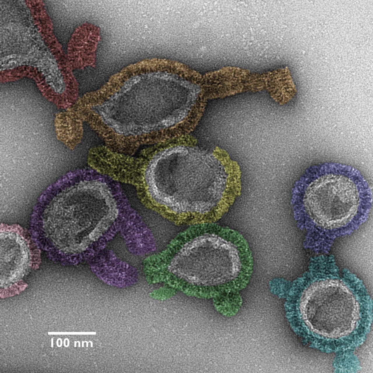
The micrograph image provides insight into the assembly pathway of antimicrobial guanylate-binding proteins (GBP1s) at a high structural resolution. It adds to our understanding of GBP-orchestrated host immunity against bacterial infection. GPB1 forms an antimicrobial protein coat on bacteria and thereby plays a critical role in host defense against bacterial pathogens.
A false-colored negative-stained transmission electron micrograph image shown at 73,000x nominal magnification, captured using a transmission electron microscope operating at 120 kV.

Olfactory bulbs of barely a day-old-mice. RNA expression of the mechanosensitive channel Piezo2 is shown in magenta, cell nuclei are marked in yellow. Note the ring-like pattern of Piezo2 expression in the two mirror-symmetric olfactory bulbs. Imaging led to identification of neurons that express Piezo2 and demonstrated that central olfactory neurons can sense mechanical stimuli.
The image was captured using an Olympus IX83 and CSU-W1 spinning disk confocal system. The mRNA of interest was highlighted with fluorophores through RNAScope, with the addition of final touches.

The dynamics of vimentin protein fibers in endothelial cell monolayers under simulated blood flow conditions. Vimentin networks’ show a morphological shift, aligning in the direction of the simulated flow, a phenomenon along with its nuclear “blanketing” effect that had not been observed before. Such flow-induced fiber alignment suggests a protective mechanism for cell nuclei.
Captured with a Zeiss LSM 980 confocal microscope using a 63x 1.4NA oil objective in SR-4Y multiplex airy scan mode for super-resolution imaging, three separate excitation/emission filters and laser configurations were utilized to image different molecular targets. These were then overlaid using differently colored lookup tables. A total of 32 z‑slices, 150 nanometers apart, were merged into a single image through Z‑Projection. Color Lookup Tables (LUTs) were applied: Red for DAPI (DNA), MPI-Inferno for VE-Cadherin antibody, and Cyan Hot for vimentin antibody immunofluorescence, to distinguish the different elements clearly.

A fruit fly embryo changes drastically in only about twelve hours:
Undifferentiated cells (2 o’clock) assume different identities (4 o’clock), migrate into individual positions (6‒10 o’clock) and produce differentiated cell types (12 o’clock), including nerve cells and muscle cells. The embryos are positioned with their heads to the left. 2 o’clock: cell nuclei (blue); 4‒10 o’clock: key genes for neuronal identity (RNA in situ hybridization, red, vnd; green, ind; blue, Dr); 12 o’clock: differentiated cell types (antibody coloring: red, cell bodies of neurons; green, axons of projection neurons; blue, muscle cells).
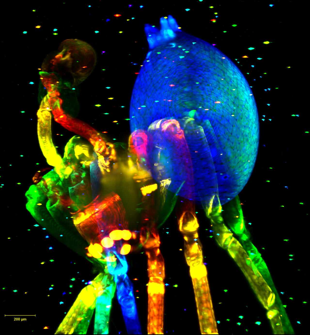
1st place 2017: Colorful spider
This is a tiny spider, about 2 mm long, that was imaged by a light sheet microscope for testing purposes. On the right is its abdomen (blue) and on the left its cephalothorax, or head (green). The spider’s mouth parts (red/yellow) can be seen underneath the cephalothorax, and its pedipalps (also red/yellow) can be seen on the front of the head. The bright dots spread throughout the whole image are caused by small fluorescent beads (see project description). The light seen here is fluorescent light and was produced by using laser-excited fluorescence to exploit the autofluorescence properties of the spider’s exoskeleton.
About the project
Researchers captured this image while assessing the imaging and analysis technology of a light sheet microscope (Lightsheet Z.1) at the MDC’s Advanced Light Microscopy (ALM) Core Facility, using a spider they just happened to find as a test specimen. It was embedded in agarose, along with fluorescent beads (measuring 6 μm in diameter) needed for spatially reconstructing the complete image – a process achieved by fusing data sets registered from eight different viewing angles of 45°. The size of the final data set was 25 GB.
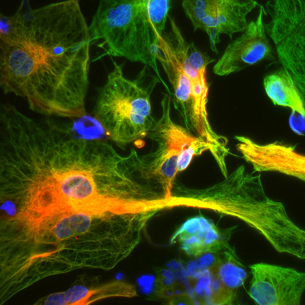
2nd place 2017: glia cells
Seen here are astrocytes (a type of glial cells) in a neuronal cell culture of Leigh syndrome patients obtained from induced pluripotent stem (iPS) cells. Glial cells, along with neurons, are an important component of the brain’s nerve tissue. The different types of cells must interact and communicate with one another so that everything functions smoothly. Immunofluorescent staining shows the proteins vimentin (green) and GFAP (rot), which are parts of the cytoskeleton and thus help to stabilize cells mechanically.
About the project
In this project, we aim to create a model of Leigh syndrome using iPS cell technology. Leigh syndrome is an early-onset, genetic multisystem disorder caused by malfunctioning mitochondria. The condition is characterized by psychomotor regression, rapidly progressing neurological symptoms, blindness, and loss of hearing as well as consciousness and respiratory disturbances. It typically results in death within a few years. We hope to find phenotypes by comparing nerves and glial cells from Leigh syndrome patients with control cell lines derived from healthy subjects.
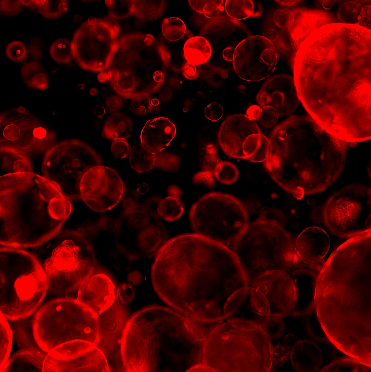
3rd place 2017: organoids of colon cancer
This is a fluorescent microscopic image showing a three-dimensional (3D) organoid culture of a metastatic colon cancer model. The tumor cells within this culture grew into balloon-like spheroids consisting of self-renewing and rapidly dividing cells. This morphology differs from organoid cultures composed of unaltered cells that create “mini colons” complete with all specialized cells. The tumor organoids shown here were further modified to produce a red fluorescent protein and to enable certain genes to be selectively switched on and off.
About the project:
Colon cancer is a common disease worldwide. Improved early detection has led to lower mortality rates, but patients with metastatic tumors still succumb to the disease. In this project, we are using an advanced 3D organoid culture as it effectively mimics the complex cell system of living organs, making it possible to investigate the efficacy of potential drugs as well as new targets for colon cancer treatment.

1st place 2016
The image is in fact a three-dimensional reconstruction of a mouse embryo made up of 360 individual images. The developing nervous system is visible in green.
Fine nerve fibers run through the embryo. The nervous system is created during embryonic development, keeps developing after birth, and remains a flexible and adaptable organ. The fine neuronal structures are extremely sensitive throughout pregnancy and vulnerable to external influences. That’s why alcohol consumption, smoking, radiation and certain maternal conditions can cause damage to the embryo’s developing nervous system.
Groups: Walter Birchmeier und Anje Sporbert


The picture shows a heart muscle precursor cell in rats. The cell nucleus has been stained blue. Two proteins involved in degrading proteins that are defective or no longer required have been labelled with a red or green fluorescing protein. They are located in the same places, so the colors are superimposed (yellow)
The degrading of proteins in cells is coordinated mainly by the ubiquitin proteasome system. A protein that is to be degraded is tagged with the small protein ubiquitin; it can then be recognized by the proteasome and degraded. E3 ubiquitin ligases play an important part in this process. They bind the protein that is to be degraded and then coordinate ubiquitin transfer. The most important E3 ubiquitin ligases in the muscle are the muscle-specific RING finger proteins (MURF). A project aimed at further characterization of the MURFs investigated, among other things, the distribution of MURF proteins in heart muscle cells.
Group: Thomas Sommer

In order to trace the organization and development of the pancreas, we observe its growth in mouse embryos from day 11.5 of embryonic development outside the body, in a dish. Here we show such an explant, in which the cell membrane is stained blue (a staining for the E‑cadherin molecule). The pink staining is the hormone insulin, which is only produced in the beta cells.
Diabetes is an incurable disease that affects more than 250 million people worldwide. Diabetics lack functional endocrine cells in the pancreas – the beta cells that excrete the hormone insulin and thereby regulate our blood sugar. Cell-based therapies offer a promising prospect of a cure for Type 1 diabetes, but they require detailed investigation of the embryonic development of the pancreas. The researcher behind the picture is studying how beta cells arise from endocrine precursor cells and how they organize themselves into three-dimensional structures, the islets of Langerhans.
Group: Francesca Spagnoli

The hippocampus is a part of the cerebrum in which memories are formed and linked. Because of its orderly structure it is a popular model for neurophysiological investigation. This picture shows a section of the hippocampus, the CA1 region. Interneurons have been labelled with an anti-ErbB4 antibody (magenta). Nrg3 has been detected with a second antibody (cyan). The overlap of ErbB4 and Nrg3 on the surface of the interneurons is indicated by the dark blue coloring.
The families of neuregulin ligands and ErbB receptors are molecules that transfer signals between various cells. Neuregulins are released by one type of cell and bind the ErbB receptors on the surface of an adjacent cell, rather like a lock and key. This binding activates the receptor, which then alters proteins in the cytoplasm of the cell enzymatically, thereby conducting the signal into the cell.
Neuregulins and ErbB receptors have important functions during development of the embryo and continue to exert an effect in the adult organism. It has been known since 2002 that certain variants of the genes that code for neuregulin‑1 and ‑3 and the receptor ErbB4 occur particularly frequently in schizophrenia patients. The research group is investigating the function of neuregulin‑3 (Nrg3) and ErbB4 in the brains of mice. The researchers have established that ErbB4 is produced by a small population of certain nerve cells, the fast-spiking interneurons. Nrg3 binds to ErbB4 on the surface of these neurons. The group is currently analyzing how Nrg3 influences the properties of the ErbB4-positive interneurons and hence the functioning of the brain.
Group: Carmen Birchmeier-Kohler

This picture shows nerve cells (pink) in the hippocampus – an area of the brain that is particularly susceptible to epilepsy. The blue coloring indicates cell nuclei in which pathogenic reworking of the protein code is taking place.
Some 50 million people worldwide suffer from chronic epilepsy. With varying degrees of frequency, sufferers experience seizures that significantly restrict their everyday lives. Epilepsy patients manage their condition with drugs, but sooner or later these unfortunately become ineffective for many. One solution is surgical removal of the affected area of the brain, the hippocampus. Joachim Meier’s group is studying certain processes at molecular level that play a part in the disease. A main issue is the fact that the nerve cells retrospectively change their prescribed code for producing protein, almost as though they were using Tipp-Ex. This results in abnormal proteins that influence the activity of the nerve cell.
Group: Jochen Meier

The picture shows a heart muscle precursor cell in the rat. The cell nucleus has been stained blue and the actin cytoskeleton green. A protein that is involved in degrading proteins that are no longer required has been labelled in yellow. It is located in small dot-like structures in the periphery of the cell nucleus.
The degrading of proteins in cells is coordinated mainly by the ubiquitin proteasome system. The protein that is to be degraded is tagged with the small protein ubiquitin; it can then be recognized by the proteasome and degraded. E3 ubiquitin ligases play an important part in this process. They bind the protein that is to be degraded and then coordinate the ubiquitin transfer. The most important E3 ubiquitin ligases in the muscle are the muscle-specific RING finger proteins (MURF). A project aimed at further characterization of the MURFs investigated, among other things, the distribution of MURF proteins in heart muscle cells.
Group: Thomas Sommer

Polyps in the intestinal mucosa are a pre-cancerous condition that can lead to bowel cancer. The formation of such polyps and of the tumor itself is triggered mainly by a Wnt signaling pathway in the cells. This picture shows a section through a human intestinal polyp with traces of healthy mucous membrane, enlarged 200 times. It uses an antibody-based staining technique (immunohistochemistry) with fluorescence labelling. Colored pink is a growth-inhibiting CCAAT/enhancer binding protein transcription factor; the green is the proto-oncoprotein beta-catenin and the blue is the cell nuclei. Cells with beta-catenin in the nucleus (light green because of the superimposition of green and blue) have an activated Wnt signaling pathway.
This fluorescent immunohistochemistry comes from a project that is investigating the carcinogenesis of colorectal cancer. The project is exploring a possible connection between the expression and tumor-suppression properties of a proliferation-inhibiting and differentiation-promoting CCAAT/enhancer binding protein transcription factor on the one hand and the Wnt signaling pathway that plays a key part in colorectal cancer and its beta-catenin signaling molecule on the other. Of particular interest are the changes in the expression strengths of the transcription factor within the carcinogenesis stages and possible correlations with the clinical behavior of the cancers.
Group: Achim Leutz

Best Scientific Image Contest
https://helmholtz-imaging.de/best-scientific-image-gallery/