© David Ausserhofer / MDC Electron Microscopy Séverine Kunz Profil Team Veröffentlichungen Nachrichten Profil Team Veröffentlichungen Nachrichten Profil Mehr Infos finden Sie auf der englischsprachigen Version unserer Webseite. Our webpage Team Leiter Dr. Severine Kunz Severine.Kunz@mdc-berlin.de +49 30 9406 – 2580 Wissenschaftler Dr. Mara-Camelia Rusu 31.2: Max-Delbrück-Haus Raum: 0218 Mara-Camelia.Rusu@mdc-berlin.de +49 30 9406 – 2578 Sekretariat Clara Pauline Lüty 87: Timoféeff-Ressovsky-Haus Raum: 1.13 ClaraPauline.Luety@mdc-berlin.de +49 30 9406 – 2884 Technischer Assistent Madlen Driesner 31.2: Max-Delbrück-Haus Raum: 0218 m.driesner@mdc-berlin.de +49 30 9406 – 3437 31.2: Max-Delbrück-Haus Raum: 0264 m.driesner@mdc-berlin.de +49 30 9406 – 3438 Christina Schiel 31.2: Max-Delbrück-Haus Raum: 0263 Christina.Schiel@mdc-berlin.de +49 30 9406 – 3438 Veröffentlichungen 22. Juni 2026 / Advanced Science Enhancing maturation of human neuromuscular organoids via electrical stimulation Chrysanthi-Maria Moysidou Inês Afonso Martins Ismail Amr El-Shimy Iacopo Bicci Donatella Cea Christina Bukas Isra Mekki Mara-Camelia Rusu Aylin Nebol Ines Lahmann Marie Piraud Enrico Klotzsch Mina Gouti Juni 2026 / Molecular Psychiatry Distinct roles for MNK1 and MNK2 in social and cognitive behavior through kinase-specific regulation of the synaptic proteome and phosphoproteome Rosalba Olga Proce Maria Steinecker Chiara Giacomelli Erika Uddström Anirban Chatterjee Souhaila Wüsthoff Luiz Gustavo Teixeira Alves Oliver Popp Tobias Pohl Katie Maxwell Lucie Hortmann Severine Kunz Philipp Mertins Markus Landthaler Daria Bunina Hanna Hörnberg 18. Januar 2026 / bioRxiv TENM4 is an essential transduction component for touch Mohammed A. Khallaf Angela Tzu-Lun Huang Letizia Dalmasso Sampurna Chakrabarti Rowena Groeneveld Yinth Andrea Bernal Sierra Jonathan Alexis Garcia-Contreras Emilie Nello Anja Schütz Valérie Bégay Alessandro Santuz Sarah Hedtrich Wei Zhong Christina Schiel Steven J. Middleton Oliver Popp Niccolò Zampieri Philipp Mertins Severine Kunz Gary R. Lewin Dezember 2025 / JACC: Advances Translational insights into myocardial deformation and fibrosis in hypertrophic cardiomyopathy using diffusion tensor MRI Oumaima Laghzali Danielle Kara Shi Chen Siqin Liu Shahriar Shalikar Mara-Camelia Rusu Fatimunnisa Qadri Lucie Carrier Hsin-Jung Yang Thoralf Niendorf Min-Chi Ku Christopher Nguyen November 2025 / Nature Metabolism Interaction of sortilin with apolipoprotein E3 enables neurons to use long-chain fatty acids as alternative metabolic fuel Anna K. Greda Jemila P. Gomes Vanessa Schmidt-Krueger Ewa Zurawska-Plaksej Raphaela Fritsche-Guenther Ina-Maria Rudolph Narasimha S. Telugu Cagla Cömert Jennifer Kirwan Séverine Kunz Michael Rothe Mogens Johannsen Sebastian Diecke Peter Bross Thomas E. Willnow 18. Juli 2025 / bioRxiv Spatiotemporal dynamics of tumor microenvironment remodeling Kamil Lisek Ilan Theurillat Tancredi Massimo Pentimalli Svea Beier Daniel León-Periñán Anna Antonatou Serafima Dubnov Marion Müller Florian Hubl Artemis Xhuri Hanna Romanowicz Beata Smolarz Elodie Montaudon Sandra Raimundo Anca Margineanu Marie Schott Séverine Kunz Elisabetta Marangoni Nikos Karaiskos Mor Nitzan Walter Birchmeier Nikolaus Rajewsky 18. Juli 2025 / bioRxiv Spatiotemporal dynamics of tumor microenvironment remodeling Kamil Lisek Ilan Theurillat Tancredi Massimo Pentimalli Svea Beier Daniel León-Periñán Anna Antonatou Serafima Dubnov Marion Müller Florian Hubl Artemis Xhuri Hanna Romanowicz Beata Smolarz Elodie Montaudon Sandra Raimundo Anca Margineanu Marie Schott Séverine Kunz Elisabetta Marangoni Nikos Karaiskos Mor Nitzan Walter Birchmeier Nikolaus Rajewsky 23. Mai 2025 / Science Advances Metabolic traits shape responses to LSD1-directed therapy in glioblastoma tumor-initiating cells G. Marotta D. Osti E. Zaccheroni B. Costanza S. Faletti A. Marinaro C. Richichi D. Mesa S. Rodighiero C. Soriani E. Migliaccio F. Ruscitto C. Priami S. Sigismund F. Manetti D. Polli G.V. Beznusenko M.C. Rusu F. Favero D. Corà D.A. Silvestris A. Gallo V. Gambino F. Alfieri S. Gandini M.J. Schmitt G. Gargiulo R. Noberini T. Bonaldi G. Pelicci 07. Februar 2025 / International Journal of Molecular Sciences Cardiac MRI strain as an early indicator of myocardial dysfunction in hypertrophic cardiomyopathy S. Liu O. Laghzali S. Shalikar M.C. Rusu L. Carrier T. Niendorf M.C. Ku 07. Februar 2025 / bioRxiv Formation of amyloid-like HTTex1 aggregates in neurons, downregulation of synaptic proteins and early mortality of Huntington’s disease flies are causally linked A. Ast L. Roth L. Brusendorf F. Schindler O.W.M. Ammar S. Berberich J. Edel M. Bonsor E. Georgii C. Hänig C. Langnick A. Ivanov D. Beule M. Piraud S. Kunz O. Popp P. Mertins A.G. Petzoldt E.E. Wanker Seitennummerierung Aktuelle Seite 1 Seite 2 Seite 3 Nächste Seite ›› Letzte Seite Last » Nachrichten Pressemitteilung Nr. 26 21. Oktober 2025 Berlin Warum ApoE4 das Risiko für Alzheimer erhöht Die Genvariante ApoE4 wird mit einem erhöhten Alzheimer-Risiko in Verbindung gebracht. Wie genau sie die neuronale Funktion im alternden Gehirn beeinträchtigt, haben jetzt Forschende des Max Delbrück Center und der Universität Aarhus entdeckt. In „Nature Metabolism“ stellen sie den Mechanismus vor. Pressemitteilung Nr. 9 29. Februar 2024 Berlin Ein neuer Kanal für Berührung Der Tastsinn ist elementar für uns – und trotzdem kaum verstanden. Jetzt hat das Team um Gary Lewin am Max Delbrück Center einen zweiten Ionenkanal entdeckt, der mit der Wahrnehmung von Berührung zu tun hat. Elkin1 könne ein Angriffspunkt für die Schmerztherapie sein, schreibt das Team in „Science“. Institut & Campus 21. Januar 2022 Auf dem Weg von 2D zu 3D Ihre Welt ist eine der Grautöne: die Elektronenmikroskopie, die Schwarz-Weiß-Aufnahmen aus dem Inneren von Zellen und Strukturen liefert. Dr. Séverine Kunz hat sich dieser verborgenen Welt verschrieben. Seit kurzem leitet sie die Technologie-Plattform Elektronenmikroskopie am MDC. Wissenschaft 26. Januar 2016 By Russell Hodge Manches muss man einfach gesehen haben Elektronenmikroskopie am MDC Dr. Severine KunzKontaktSeverine.Kunz@mdc-berlin.deTelefon: +49 30 9406-2580Max-Delbrück-Centrum für Molekulare Medizin (MDC)Robert-Rössle-Straße 1013125 Berlin, DeutschlandHaus 31.2, Raum 1214.1
© David Ausserhofer / MDC Electron Microscopy Séverine Kunz Profil Team Veröffentlichungen Nachrichten Profil Team Veröffentlichungen Nachrichten Profil Mehr Infos finden Sie auf der englischsprachigen Version unserer Webseite. Our webpage Team Leiter Dr. Severine Kunz Severine.Kunz@mdc-berlin.de +49 30 9406 – 2580 Wissenschaftler Dr. Mara-Camelia Rusu 31.2: Max-Delbrück-Haus Raum: 0218 Mara-Camelia.Rusu@mdc-berlin.de +49 30 9406 – 2578 Sekretariat Clara Pauline Lüty 87: Timoféeff-Ressovsky-Haus Raum: 1.13 ClaraPauline.Luety@mdc-berlin.de +49 30 9406 – 2884 Technischer Assistent Madlen Driesner 31.2: Max-Delbrück-Haus Raum: 0218 m.driesner@mdc-berlin.de +49 30 9406 – 3437 31.2: Max-Delbrück-Haus Raum: 0264 m.driesner@mdc-berlin.de +49 30 9406 – 3438 Christina Schiel 31.2: Max-Delbrück-Haus Raum: 0263 Christina.Schiel@mdc-berlin.de +49 30 9406 – 3438 Veröffentlichungen 22. Juni 2026 / Advanced Science Enhancing maturation of human neuromuscular organoids via electrical stimulation Chrysanthi-Maria Moysidou Inês Afonso Martins Ismail Amr El-Shimy Iacopo Bicci Donatella Cea Christina Bukas Isra Mekki Mara-Camelia Rusu Aylin Nebol Ines Lahmann Marie Piraud Enrico Klotzsch Mina Gouti Juni 2026 / Molecular Psychiatry Distinct roles for MNK1 and MNK2 in social and cognitive behavior through kinase-specific regulation of the synaptic proteome and phosphoproteome Rosalba Olga Proce Maria Steinecker Chiara Giacomelli Erika Uddström Anirban Chatterjee Souhaila Wüsthoff Luiz Gustavo Teixeira Alves Oliver Popp Tobias Pohl Katie Maxwell Lucie Hortmann Severine Kunz Philipp Mertins Markus Landthaler Daria Bunina Hanna Hörnberg 18. Januar 2026 / bioRxiv TENM4 is an essential transduction component for touch Mohammed A. Khallaf Angela Tzu-Lun Huang Letizia Dalmasso Sampurna Chakrabarti Rowena Groeneveld Yinth Andrea Bernal Sierra Jonathan Alexis Garcia-Contreras Emilie Nello Anja Schütz Valérie Bégay Alessandro Santuz Sarah Hedtrich Wei Zhong Christina Schiel Steven J. Middleton Oliver Popp Niccolò Zampieri Philipp Mertins Severine Kunz Gary R. Lewin Dezember 2025 / JACC: Advances Translational insights into myocardial deformation and fibrosis in hypertrophic cardiomyopathy using diffusion tensor MRI Oumaima Laghzali Danielle Kara Shi Chen Siqin Liu Shahriar Shalikar Mara-Camelia Rusu Fatimunnisa Qadri Lucie Carrier Hsin-Jung Yang Thoralf Niendorf Min-Chi Ku Christopher Nguyen November 2025 / Nature Metabolism Interaction of sortilin with apolipoprotein E3 enables neurons to use long-chain fatty acids as alternative metabolic fuel Anna K. Greda Jemila P. Gomes Vanessa Schmidt-Krueger Ewa Zurawska-Plaksej Raphaela Fritsche-Guenther Ina-Maria Rudolph Narasimha S. Telugu Cagla Cömert Jennifer Kirwan Séverine Kunz Michael Rothe Mogens Johannsen Sebastian Diecke Peter Bross Thomas E. Willnow 18. Juli 2025 / bioRxiv Spatiotemporal dynamics of tumor microenvironment remodeling Kamil Lisek Ilan Theurillat Tancredi Massimo Pentimalli Svea Beier Daniel León-Periñán Anna Antonatou Serafima Dubnov Marion Müller Florian Hubl Artemis Xhuri Hanna Romanowicz Beata Smolarz Elodie Montaudon Sandra Raimundo Anca Margineanu Marie Schott Séverine Kunz Elisabetta Marangoni Nikos Karaiskos Mor Nitzan Walter Birchmeier Nikolaus Rajewsky 18. Juli 2025 / bioRxiv Spatiotemporal dynamics of tumor microenvironment remodeling Kamil Lisek Ilan Theurillat Tancredi Massimo Pentimalli Svea Beier Daniel León-Periñán Anna Antonatou Serafima Dubnov Marion Müller Florian Hubl Artemis Xhuri Hanna Romanowicz Beata Smolarz Elodie Montaudon Sandra Raimundo Anca Margineanu Marie Schott Séverine Kunz Elisabetta Marangoni Nikos Karaiskos Mor Nitzan Walter Birchmeier Nikolaus Rajewsky 23. Mai 2025 / Science Advances Metabolic traits shape responses to LSD1-directed therapy in glioblastoma tumor-initiating cells G. Marotta D. Osti E. Zaccheroni B. Costanza S. Faletti A. Marinaro C. Richichi D. Mesa S. Rodighiero C. Soriani E. Migliaccio F. Ruscitto C. Priami S. Sigismund F. Manetti D. Polli G.V. Beznusenko M.C. Rusu F. Favero D. Corà D.A. Silvestris A. Gallo V. Gambino F. Alfieri S. Gandini M.J. Schmitt G. Gargiulo R. Noberini T. Bonaldi G. Pelicci 07. Februar 2025 / International Journal of Molecular Sciences Cardiac MRI strain as an early indicator of myocardial dysfunction in hypertrophic cardiomyopathy S. Liu O. Laghzali S. Shalikar M.C. Rusu L. Carrier T. Niendorf M.C. Ku 07. Februar 2025 / bioRxiv Formation of amyloid-like HTTex1 aggregates in neurons, downregulation of synaptic proteins and early mortality of Huntington’s disease flies are causally linked A. Ast L. Roth L. Brusendorf F. Schindler O.W.M. Ammar S. Berberich J. Edel M. Bonsor E. Georgii C. Hänig C. Langnick A. Ivanov D. Beule M. Piraud S. Kunz O. Popp P. Mertins A.G. Petzoldt E.E. Wanker Seitennummerierung Aktuelle Seite 1 Seite 2 Seite 3 Nächste Seite ›› Letzte Seite Last » Nachrichten Pressemitteilung Nr. 26 21. Oktober 2025 Berlin Warum ApoE4 das Risiko für Alzheimer erhöht Die Genvariante ApoE4 wird mit einem erhöhten Alzheimer-Risiko in Verbindung gebracht. Wie genau sie die neuronale Funktion im alternden Gehirn beeinträchtigt, haben jetzt Forschende des Max Delbrück Center und der Universität Aarhus entdeckt. In „Nature Metabolism“ stellen sie den Mechanismus vor. Pressemitteilung Nr. 9 29. Februar 2024 Berlin Ein neuer Kanal für Berührung Der Tastsinn ist elementar für uns – und trotzdem kaum verstanden. Jetzt hat das Team um Gary Lewin am Max Delbrück Center einen zweiten Ionenkanal entdeckt, der mit der Wahrnehmung von Berührung zu tun hat. Elkin1 könne ein Angriffspunkt für die Schmerztherapie sein, schreibt das Team in „Science“. Institut & Campus 21. Januar 2022 Auf dem Weg von 2D zu 3D Ihre Welt ist eine der Grautöne: die Elektronenmikroskopie, die Schwarz-Weiß-Aufnahmen aus dem Inneren von Zellen und Strukturen liefert. Dr. Séverine Kunz hat sich dieser verborgenen Welt verschrieben. Seit kurzem leitet sie die Technologie-Plattform Elektronenmikroskopie am MDC. Wissenschaft 26. Januar 2016 By Russell Hodge Manches muss man einfach gesehen haben Elektronenmikroskopie am MDC Dr. Severine KunzKontaktSeverine.Kunz@mdc-berlin.deTelefon: +49 30 9406-2580Max-Delbrück-Centrum für Molekulare Medizin (MDC)Robert-Rössle-Straße 1013125 Berlin, DeutschlandHaus 31.2, Raum 1214.1
Team Leiter Dr. Severine Kunz Severine.Kunz@mdc-berlin.de +49 30 9406 – 2580 Wissenschaftler Dr. Mara-Camelia Rusu 31.2: Max-Delbrück-Haus Raum: 0218 Mara-Camelia.Rusu@mdc-berlin.de +49 30 9406 – 2578 Sekretariat Clara Pauline Lüty 87: Timoféeff-Ressovsky-Haus Raum: 1.13 ClaraPauline.Luety@mdc-berlin.de +49 30 9406 – 2884 Technischer Assistent Madlen Driesner 31.2: Max-Delbrück-Haus Raum: 0218 m.driesner@mdc-berlin.de +49 30 9406 – 3437 31.2: Max-Delbrück-Haus Raum: 0264 m.driesner@mdc-berlin.de +49 30 9406 – 3438 Christina Schiel 31.2: Max-Delbrück-Haus Raum: 0263 Christina.Schiel@mdc-berlin.de +49 30 9406 – 3438
Veröffentlichungen 22. Juni 2026 / Advanced Science Enhancing maturation of human neuromuscular organoids via electrical stimulation Chrysanthi-Maria Moysidou Inês Afonso Martins Ismail Amr El-Shimy Iacopo Bicci Donatella Cea Christina Bukas Isra Mekki Mara-Camelia Rusu Aylin Nebol Ines Lahmann Marie Piraud Enrico Klotzsch Mina Gouti Juni 2026 / Molecular Psychiatry Distinct roles for MNK1 and MNK2 in social and cognitive behavior through kinase-specific regulation of the synaptic proteome and phosphoproteome Rosalba Olga Proce Maria Steinecker Chiara Giacomelli Erika Uddström Anirban Chatterjee Souhaila Wüsthoff Luiz Gustavo Teixeira Alves Oliver Popp Tobias Pohl Katie Maxwell Lucie Hortmann Severine Kunz Philipp Mertins Markus Landthaler Daria Bunina Hanna Hörnberg 18. Januar 2026 / bioRxiv TENM4 is an essential transduction component for touch Mohammed A. Khallaf Angela Tzu-Lun Huang Letizia Dalmasso Sampurna Chakrabarti Rowena Groeneveld Yinth Andrea Bernal Sierra Jonathan Alexis Garcia-Contreras Emilie Nello Anja Schütz Valérie Bégay Alessandro Santuz Sarah Hedtrich Wei Zhong Christina Schiel Steven J. Middleton Oliver Popp Niccolò Zampieri Philipp Mertins Severine Kunz Gary R. Lewin Dezember 2025 / JACC: Advances Translational insights into myocardial deformation and fibrosis in hypertrophic cardiomyopathy using diffusion tensor MRI Oumaima Laghzali Danielle Kara Shi Chen Siqin Liu Shahriar Shalikar Mara-Camelia Rusu Fatimunnisa Qadri Lucie Carrier Hsin-Jung Yang Thoralf Niendorf Min-Chi Ku Christopher Nguyen November 2025 / Nature Metabolism Interaction of sortilin with apolipoprotein E3 enables neurons to use long-chain fatty acids as alternative metabolic fuel Anna K. Greda Jemila P. Gomes Vanessa Schmidt-Krueger Ewa Zurawska-Plaksej Raphaela Fritsche-Guenther Ina-Maria Rudolph Narasimha S. Telugu Cagla Cömert Jennifer Kirwan Séverine Kunz Michael Rothe Mogens Johannsen Sebastian Diecke Peter Bross Thomas E. Willnow 18. Juli 2025 / bioRxiv Spatiotemporal dynamics of tumor microenvironment remodeling Kamil Lisek Ilan Theurillat Tancredi Massimo Pentimalli Svea Beier Daniel León-Periñán Anna Antonatou Serafima Dubnov Marion Müller Florian Hubl Artemis Xhuri Hanna Romanowicz Beata Smolarz Elodie Montaudon Sandra Raimundo Anca Margineanu Marie Schott Séverine Kunz Elisabetta Marangoni Nikos Karaiskos Mor Nitzan Walter Birchmeier Nikolaus Rajewsky 18. Juli 2025 / bioRxiv Spatiotemporal dynamics of tumor microenvironment remodeling Kamil Lisek Ilan Theurillat Tancredi Massimo Pentimalli Svea Beier Daniel León-Periñán Anna Antonatou Serafima Dubnov Marion Müller Florian Hubl Artemis Xhuri Hanna Romanowicz Beata Smolarz Elodie Montaudon Sandra Raimundo Anca Margineanu Marie Schott Séverine Kunz Elisabetta Marangoni Nikos Karaiskos Mor Nitzan Walter Birchmeier Nikolaus Rajewsky 23. Mai 2025 / Science Advances Metabolic traits shape responses to LSD1-directed therapy in glioblastoma tumor-initiating cells G. Marotta D. Osti E. Zaccheroni B. Costanza S. Faletti A. Marinaro C. Richichi D. Mesa S. Rodighiero C. Soriani E. Migliaccio F. Ruscitto C. Priami S. Sigismund F. Manetti D. Polli G.V. Beznusenko M.C. Rusu F. Favero D. Corà D.A. Silvestris A. Gallo V. Gambino F. Alfieri S. Gandini M.J. Schmitt G. Gargiulo R. Noberini T. Bonaldi G. Pelicci 07. Februar 2025 / International Journal of Molecular Sciences Cardiac MRI strain as an early indicator of myocardial dysfunction in hypertrophic cardiomyopathy S. Liu O. Laghzali S. Shalikar M.C. Rusu L. Carrier T. Niendorf M.C. Ku 07. Februar 2025 / bioRxiv Formation of amyloid-like HTTex1 aggregates in neurons, downregulation of synaptic proteins and early mortality of Huntington’s disease flies are causally linked A. Ast L. Roth L. Brusendorf F. Schindler O.W.M. Ammar S. Berberich J. Edel M. Bonsor E. Georgii C. Hänig C. Langnick A. Ivanov D. Beule M. Piraud S. Kunz O. Popp P. Mertins A.G. Petzoldt E.E. Wanker Seitennummerierung Aktuelle Seite 1 Seite 2 Seite 3 Nächste Seite ›› Letzte Seite Last »
22. Juni 2026 / Advanced Science Enhancing maturation of human neuromuscular organoids via electrical stimulation Chrysanthi-Maria Moysidou Inês Afonso Martins Ismail Amr El-Shimy Iacopo Bicci Donatella Cea Christina Bukas Isra Mekki Mara-Camelia Rusu Aylin Nebol Ines Lahmann Marie Piraud Enrico Klotzsch Mina Gouti
Juni 2026 / Molecular Psychiatry Distinct roles for MNK1 and MNK2 in social and cognitive behavior through kinase-specific regulation of the synaptic proteome and phosphoproteome Rosalba Olga Proce Maria Steinecker Chiara Giacomelli Erika Uddström Anirban Chatterjee Souhaila Wüsthoff Luiz Gustavo Teixeira Alves Oliver Popp Tobias Pohl Katie Maxwell Lucie Hortmann Severine Kunz Philipp Mertins Markus Landthaler Daria Bunina Hanna Hörnberg
18. Januar 2026 / bioRxiv TENM4 is an essential transduction component for touch Mohammed A. Khallaf Angela Tzu-Lun Huang Letizia Dalmasso Sampurna Chakrabarti Rowena Groeneveld Yinth Andrea Bernal Sierra Jonathan Alexis Garcia-Contreras Emilie Nello Anja Schütz Valérie Bégay Alessandro Santuz Sarah Hedtrich Wei Zhong Christina Schiel Steven J. Middleton Oliver Popp Niccolò Zampieri Philipp Mertins Severine Kunz Gary R. Lewin
Dezember 2025 / JACC: Advances Translational insights into myocardial deformation and fibrosis in hypertrophic cardiomyopathy using diffusion tensor MRI Oumaima Laghzali Danielle Kara Shi Chen Siqin Liu Shahriar Shalikar Mara-Camelia Rusu Fatimunnisa Qadri Lucie Carrier Hsin-Jung Yang Thoralf Niendorf Min-Chi Ku Christopher Nguyen
November 2025 / Nature Metabolism Interaction of sortilin with apolipoprotein E3 enables neurons to use long-chain fatty acids as alternative metabolic fuel Anna K. Greda Jemila P. Gomes Vanessa Schmidt-Krueger Ewa Zurawska-Plaksej Raphaela Fritsche-Guenther Ina-Maria Rudolph Narasimha S. Telugu Cagla Cömert Jennifer Kirwan Séverine Kunz Michael Rothe Mogens Johannsen Sebastian Diecke Peter Bross Thomas E. Willnow
18. Juli 2025 / bioRxiv Spatiotemporal dynamics of tumor microenvironment remodeling Kamil Lisek Ilan Theurillat Tancredi Massimo Pentimalli Svea Beier Daniel León-Periñán Anna Antonatou Serafima Dubnov Marion Müller Florian Hubl Artemis Xhuri Hanna Romanowicz Beata Smolarz Elodie Montaudon Sandra Raimundo Anca Margineanu Marie Schott Séverine Kunz Elisabetta Marangoni Nikos Karaiskos Mor Nitzan Walter Birchmeier Nikolaus Rajewsky
18. Juli 2025 / bioRxiv Spatiotemporal dynamics of tumor microenvironment remodeling Kamil Lisek Ilan Theurillat Tancredi Massimo Pentimalli Svea Beier Daniel León-Periñán Anna Antonatou Serafima Dubnov Marion Müller Florian Hubl Artemis Xhuri Hanna Romanowicz Beata Smolarz Elodie Montaudon Sandra Raimundo Anca Margineanu Marie Schott Séverine Kunz Elisabetta Marangoni Nikos Karaiskos Mor Nitzan Walter Birchmeier Nikolaus Rajewsky
23. Mai 2025 / Science Advances Metabolic traits shape responses to LSD1-directed therapy in glioblastoma tumor-initiating cells G. Marotta D. Osti E. Zaccheroni B. Costanza S. Faletti A. Marinaro C. Richichi D. Mesa S. Rodighiero C. Soriani E. Migliaccio F. Ruscitto C. Priami S. Sigismund F. Manetti D. Polli G.V. Beznusenko M.C. Rusu F. Favero D. Corà D.A. Silvestris A. Gallo V. Gambino F. Alfieri S. Gandini M.J. Schmitt G. Gargiulo R. Noberini T. Bonaldi G. Pelicci
07. Februar 2025 / International Journal of Molecular Sciences Cardiac MRI strain as an early indicator of myocardial dysfunction in hypertrophic cardiomyopathy S. Liu O. Laghzali S. Shalikar M.C. Rusu L. Carrier T. Niendorf M.C. Ku
07. Februar 2025 / bioRxiv Formation of amyloid-like HTTex1 aggregates in neurons, downregulation of synaptic proteins and early mortality of Huntington’s disease flies are causally linked A. Ast L. Roth L. Brusendorf F. Schindler O.W.M. Ammar S. Berberich J. Edel M. Bonsor E. Georgii C. Hänig C. Langnick A. Ivanov D. Beule M. Piraud S. Kunz O. Popp P. Mertins A.G. Petzoldt E.E. Wanker
Nachrichten Pressemitteilung Nr. 26 21. Oktober 2025 Berlin Warum ApoE4 das Risiko für Alzheimer erhöht Die Genvariante ApoE4 wird mit einem erhöhten Alzheimer-Risiko in Verbindung gebracht. Wie genau sie die neuronale Funktion im alternden Gehirn beeinträchtigt, haben jetzt Forschende des Max Delbrück Center und der Universität Aarhus entdeckt. In „Nature Metabolism“ stellen sie den Mechanismus vor. Pressemitteilung Nr. 9 29. Februar 2024 Berlin Ein neuer Kanal für Berührung Der Tastsinn ist elementar für uns – und trotzdem kaum verstanden. Jetzt hat das Team um Gary Lewin am Max Delbrück Center einen zweiten Ionenkanal entdeckt, der mit der Wahrnehmung von Berührung zu tun hat. Elkin1 könne ein Angriffspunkt für die Schmerztherapie sein, schreibt das Team in „Science“. Institut & Campus 21. Januar 2022 Auf dem Weg von 2D zu 3D Ihre Welt ist eine der Grautöne: die Elektronenmikroskopie, die Schwarz-Weiß-Aufnahmen aus dem Inneren von Zellen und Strukturen liefert. Dr. Séverine Kunz hat sich dieser verborgenen Welt verschrieben. Seit kurzem leitet sie die Technologie-Plattform Elektronenmikroskopie am MDC. Wissenschaft 26. Januar 2016 By Russell Hodge Manches muss man einfach gesehen haben Elektronenmikroskopie am MDC
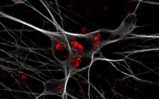
Pressemitteilung Nr. 26 21. Oktober 2025 Berlin Warum ApoE4 das Risiko für Alzheimer erhöht Die Genvariante ApoE4 wird mit einem erhöhten Alzheimer-Risiko in Verbindung gebracht. Wie genau sie die neuronale Funktion im alternden Gehirn beeinträchtigt, haben jetzt Forschende des Max Delbrück Center und der Universität Aarhus entdeckt. In „Nature Metabolism“ stellen sie den Mechanismus vor.
Pressemitteilung Nr. 9 29. Februar 2024 Berlin Ein neuer Kanal für Berührung Der Tastsinn ist elementar für uns – und trotzdem kaum verstanden. Jetzt hat das Team um Gary Lewin am Max Delbrück Center einen zweiten Ionenkanal entdeckt, der mit der Wahrnehmung von Berührung zu tun hat. Elkin1 könne ein Angriffspunkt für die Schmerztherapie sein, schreibt das Team in „Science“.
Institut & Campus 21. Januar 2022 Auf dem Weg von 2D zu 3D Ihre Welt ist eine der Grautöne: die Elektronenmikroskopie, die Schwarz-Weiß-Aufnahmen aus dem Inneren von Zellen und Strukturen liefert. Dr. Séverine Kunz hat sich dieser verborgenen Welt verschrieben. Seit kurzem leitet sie die Technologie-Plattform Elektronenmikroskopie am MDC.
Wissenschaft 26. Januar 2016 By Russell Hodge Manches muss man einfach gesehen haben Elektronenmikroskopie am MDC
Dr. Severine KunzKontaktSeverine.Kunz@mdc-berlin.deTelefon: +49 30 9406-2580Max-Delbrück-Centrum für Molekulare Medizin (MDC)Robert-Rössle-Straße 1013125 Berlin, DeutschlandHaus 31.2, Raum 1214.1