Asymmetrical jogging and a left-handed heart
On the outside, our bodies are highly symmetrical from left to right. This mirror-image structure arises as cells in a developing embryo activate the same genes on both sides to stimulate growth and development. But a look inside reveals a different picture: Most internal organs develop asymmetrically. Nearly everyone’s heart, for example lies slightly to the left within the chest, and its left side is larger than the right. This asymmetry is a feature of the healthy organ, and even a minor disruption in the activity of genes that produce it can lead to serious heart defects. The process happens a bit differently in various animal species. All of their hearts begin as a symmetric tube of cells. In fish, the left side expands when developing cardiac cells preferentially migrate toward the left – a process known as cardiac jogging. Salim Seyfried’s group has now discovered that a complex interplay of signaling molecules is required to produce this healthy asymmetry. The current study, published in the March 25 edition of Developmental Cell, demonstrates that it arises through an unexpected type of crosstalk between important molecular signals called Nodal and Bmp.
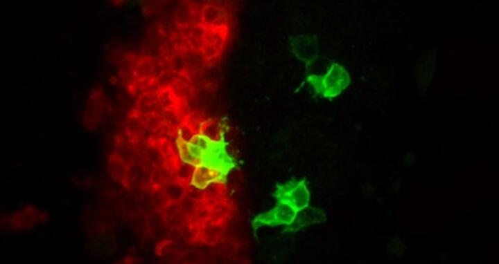
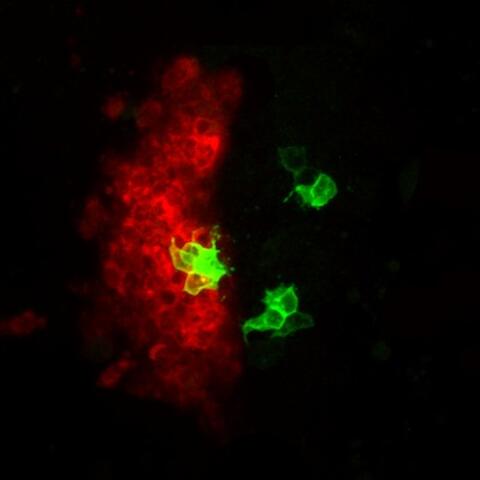
Inhibiting Bmp activity affects the asymmetric development of the heart independently of Nodal. Here the left side of the heart expresses molecules such as lefty1 (red), a sign that Nodal signaling is active. On the right side appear clusters of cells in which Bmp has been switched off (green). Within these clones, Nodal is not activated
Justus Veerkamp and other members of Salim’s lab have been studying the signals that guide heart development in the zebrafish. This animal is uniquely suited for studies of the heart. “Its embryos are virtually transparent, which means we can observe the growth of internal organs in living animals,” Salim says. “We have advanced techniques to manipulate zebrafish genes and study their effects on the development.of the heart and other organs. The embryos are so small that even with serious heart defects, they survive longer than other animals; oxygen and nutrients can simply diffuse through tissues to reach cells.”
During development, the small protein Nodal is secreted by cells and docks onto receptor molecules on the surfaces of other cells. This binding activates a signaling pathway in the target cells, changing the activity of genes and altering the cells’ behavior. Understanding Nodal’s activity in the heart requires unraveling the consequences of these complex signaling pathways within cardiac cells.
Scientists had already identified some of the molecules required to create the left/right asymmetry of the heart, including Nodal. Evolution has given zebrafish three copies that have taken on different functions in various tissues. The form of Nodal produced to the left of the zebrafish heart is called Southpaw. Researchers also knew that a second molecular signaling pathway called Bmp was involved.
“During cardiac development in zebrafish, the cells that will make up the heart take on different states,” Salim says. “Some are in a relatively immobile state, well-attached to their neighbors. Others are more mobile. We discovered that this difference turns out to have an essential effect on heart asymmetry. And as it turns out, explaining how such different states are generated within the heart required understanding the connection between Nodal and Bmp signaling.”
Work by other labs had suggested that both Nodal and Bmp might be more active on the left side of the heart. Somehow this made developing cardiac cells more mobile. Higher motility is something you would expect on the left side, Salim says, because the heart expands in that direction.
But this picture was somewhat difficult to reconcile with what the group found when they took a close look at Bmp’s presence and activity in heart tissue. First they surveyed the zebrafish heart at various stages to find out where Bmp was being produced. “Interestingly, in the phases leading up to the development of asymmetry, Bmp was produced at fairly equal levels on the left and right sides of the ‘midline’ that separates them,” Salim says.
The presence of Bmp alone, however, did not mean that it was actively passing along signals that would trigger its target genes. Here the scientists noticed an important difference: Bmp was less active on the left side. Something was dampening its signaling activity.
Nodal/Southpaw was present on the left – could it be responsible? The scientists used a method to block the Southpaw signal. This led to an increase in Bmp activity on the left side compared to normal levels. “We took this to confirm our hypothesis that under normal circumstances, Nodal was tuning down Bmp on that side,” Salim says.
In another experiment, the lab examined zebrafish that produced Southpaw on the right-hand side. In these fish Bmp activity was lower on that side, and developing cells began moving more freely there. The aberrant signals from Southpaw had reversed the asymmetry of the heart and also the activity of Bmp. To confirm this picture, the scientists interfered directly with the Bmp signal on the right side of normal zebrafish. This too flipped the growth of the heart.
“When Bmp is highly active, cells adhere more strongly and can’t migrate as freely,” Salim says. “Where it is weaker, you have higher motility and more rapid expansion. This expansion of the cardiac tube during ‘cardiac jogging’ is invariantly biased toward the left under normal physiological conditions. We can trace this to the biophysical properties of cells that move more freely on the one side, thus determining asymmetry during heart morphogenesis.”
How does Nodal block Bmp signaling on the left? The connection turned out to lie with an enzyme called Has2. Its activity also interferes with Bmp signaling. “Our hypothesis is that when Bmp molecules are secreted by the cardiac cells, they get trapped in the matrix,” Salim says. “This weakens their activity and thereby increases cardiac cell migration specifically within the left cardiac field. This faster movement apparently establishes the crucial difference between the two sides during heart morphogenesis.”
The lab used computer simulations to demonstrate that the ability of cells to “walk” in random directions – but moving slightly faster when they were positioned on the left side_– sufficed to bias heart growth.
“This work not only changes the standard picture of the activity of Nodal and Bmp signals in heart development,” Salim says, “but it also establishes the link between the two pathways. It shows that asymmetry can be traced back to the biophysical properties of cells, which can engage in a faster random movement on the left side. One result is tissue expansion in that direction.
“It also demonstrates an important role for the extracellular matrix in this process. This may help explain a number of defects in cardiac development and disease that have been linked to the matrix. There is also a potential link to cancer – in which cells detach themselves from tumors and go for ‘walks’ that turn into metastases. This work gives us a new handle on the study of the connection between the intensity of various signals and the biophysical properties of cells, and the roles that these processes play in development and disease.”
- Russ Hodge
Highlight Reference:
Veerkamp J, Rudolph F, Cseresnyes Z, Priller F, Otten C, Renz M, Schaefer L, Abdelilah-Seyfried S. Unilateral dampening of Bmp activity by nodal generates cardiac left-right asymmetry. Dev Cell. 2013 Mar 25;24(6):660-7.


