Enzyme prisons
There are up to a hundred different receptors on the surface of each cell in the human body. The cell uses these receptors to receive extracellular signals, which it then transmits to its interior. Such signals arrive at the cell in various forms, including as sensory perceptions, neurotransmitters like dopamine, or hormones like insulin.
But since the early 1980s we have known, for example, that two different heart cell receptors release exactly the same amount of cAMP when they receive an external signal, yet completely different effects are produced inside the cell.
One of the most important signaling molecules the cell uses to transmit such stimuli to its interior, which then triggers the corresponding signaling pathways, is a small molecule called cAMP. This so-called second messenger was discovered in the 1950s. Until now, experimental observations have assumed that cAMP diffuses freely – i.e., that its concentration is basically the same throughout the cell – and that one signal should therefore encompass the entire cell.
“But since the early 1980s we have known, for example, that two different heart cell receptors release exactly the same amount of cAMP when they receive an external signal, yet completely different effects are produced inside the cell,” reports Dr. Andreas Bock. Together with Dr. Paolo Annibale, Bock is temporarily heading the Receptor Signaling Lab at the Max Delbrück Center for Molecular Medicine in the Helmholtz Association (MDC) in Berlin.
Like holes in a Swiss cheese
Bock and Annibale, who are the study’s two lead authors, have now solved this apparent contradiction – which has preoccupied scientists for almost forty years. The team now reports in Cell that, contrary to previous assumptions, the majority of cAMP molecules cannot move around freely in the cell, but are actually bound to certain proteins – particularly protein kinases. In addition to the three scientists and Professor Martin Falcke from the MDC, the research project involved other Berlin researchers as well as scientists from Würzburg and Minneapolis.
“Due to this protein binding, the concentration of free cAMP in the cell is actually very low,” says Professor Martin Lohse, who is last author of the study and former head of the group. “This gives the rather slow cAMP-degrading enzymes, the phosphodiesterases (PDEs), enough time to form nanometer-sized compartments around themselves that are almost free of cAMP.” The signaling molecule is then regulated separately in each of these tiny compartments. “This enables cells to process different receptor signals simultaneously in many such compartments,” explains Lohse. The researchers were able to demonstrate this using the example of the cAMP-dependent protein kinase A (PKA), the activation of which in different compartments required different amounts of cAMP.
You can imagine these cleared-out compartments rather like the holes in a Swiss cheese – or like tiny prisons in which the actually rather slow-working PDE keeps watch over the much faster cAMP to make sure it does not break out and trigger unintended effects in the cell.
“You can imagine these cleared-out compartments rather like the holes in a Swiss cheese – or like tiny prisons in which the actually rather slow-working PDE keeps watch over the much faster cAMP to make sure it does not break out and trigger unintended effects in the cell,” explains Annibale. “Once the perpetrator is locked up, the police no longer have to chase after it.”
Nanometer-scale measurements
The team identified the movements of the signaling molecule in the cell using fluorescent cAMP molecules and special methods of fluorescence spectroscopy – including fluctuation spectroscopy and anisotropy – which Annibale developed even further for the study. So-called nanorulers helped the group to measure the size of the holes in which cAMP switches on specific signaling pathways. “These are elongated proteins that we were able to use like a tiny ruler,” explains Bock, who invented this particular nanoruler.
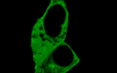
The confocal image shows cells expressing one of the newly designed nanorulers to map nanometer-size cAMP gradients in intact cells.
The team’s measurements showed that most compartments are actually smaller than 10 nanometers – i.e., 10 millionths of a millimeter. This way, the cell is able to create thousands of distinct cellular domains in which it can regulate cAMP separately and thus protect itself from the signaling molecule’s unintended effects. “We were able to show that a specific signaling pathway was initially interrupted in a hole that was virtually cAMP-free,” said Annibale. “But when we inhibited the PDEs that create these holes, the pathway continued on unobstructed.”
A chip rather than a switch
“This means the cell does not act like a single on/off switch, but rather like an entire chip containing thousands of such switches,” explains Lohse, summarizing the findings of the research. “The mistake made in past experiments was to use cAMP concentrations that were far too high, thus enabling a large amount of the signaling molecule to diffuse freely in the cell because all binding sites were occupied.”
This means the cell does not act like a single on/off switch, but rather like an entire chip containing thousands of such switches.
As a next step, the researchers want to further investigate the architecture of the cAMP “prisons” and find out which PDEs protect which signaling proteins. In the future, medical research could also benefit from their findings. “Many drugs work by altering signaling pathways within the cell,” explains Lohse. “Thanks to the discovery of this cell compartmentalization, we now know there are a great many more potential targets that can be searched for.”
“A study from San Diego, which was published at the same time as our article in Cell, shows that cells begin to proliferate when their individual signaling pathways are no longer regulated by spatial separation,” says Bock. In addition, he adds, it is already known that the distribution of cAMP concentration levels in heart cells changes in heart failure, for example. Their work could therefore open up new avenues for both cancer and cardiovascular research.
Text: Anke Brodmerkel
Further information
Grants for new research projects
Literature
Annibale, Paolo & Bock, Andreas et al. (2020): "Optical mapping of cAMP signaling at the nanometer scale"; Cell, DOI: 10.1016/j.cell.2020.07.035
Downloads
The confocal image shows cells expressing one of the newly designed nanorulers to map nanometer-size cAMP gradients in intact cells. Foto: Paolo Annibale, MDC
The slow-moving ships on the open sea serve to illustrate the limited cAMP dynamics. The whirlpools represent cAMP nanodomains around PDEs. Photo: Charlotte Konrad
Press contacts
Dr. Andreas Bock
Co-head of the Receptor Signaling Lab
Max Delbrück Center for Molecular Medicine in the Helmholtz Association (MDC)
+49-(0)30-9406-1745
Andreas.Bock@mdc-berlin.de
Professor Martin Lohse
ISAR Bioscience Institute, München
Martin.Lohse@isarbioscience.de
Christina Anders
Editor, Communications Department
Max Delbrück Center for Molecular Medicine in the Helmholtz Association (MDC)
+49-(0)30-9406-2118
christina.anders@mdc-berlin.de or presse@mdc-berlin.de
- The Max Delbrück Center for Molecular Medicine (MDC)
-
The Max Delbrück Center for Molecular Medicine in the Helmholtz Association (MDC) is one of the world’s leading biomedical research institutions. Max Delbrück, a Berlin native, was a Nobel laureate and one of the founders of molecular biology. At the MDC’s locations in Berlin-Buch and Mitte, researchers from some 60 countries analyze the human system – investigating the biological foundations of life from its most elementary building blocks to systems-wide mechanisms. By understanding what regulates or disrupts the dynamic equilibrium in a cell, an organ, or the entire body, we can prevent diseases, diagnose them earlier, and stop their progression with tailored therapies. Patients should benefit as soon as possible from basic research discoveries. The MDC therefore supports spin-off creation and participates in collaborative networks. It works in close partnership with Charité – Universitätsmedizin Berlin in the jointly run Experimental and Clinical Research Center (ECRC), the Berlin Institute of Health (BIH) at Charité, and the German Center for Cardiovascular Research (DZHK). Founded in 1992, the MDC today employs 1,600 people and is funded 90 percent by the German federal government and 10 percent by the State of Berlin.


