
The mysterious sensory system of the spinal cord
Whether it is a matter of securing food and drinking water, fleeing from potential predators, maintaining balance or engaging in sexual reproduction, nearly all animals rely on complex locomotor patterns to survive – and must constantly adapt them to external conditions. Fine-tuning requires the integration of different sensory information that is then transmitted to motor neurons controlling muscle activity.
Long known, but hardly studied

Katrin Gerstmann and Niccolò Zampieri
Sensory cells of the eyes and skin, for instance, have been well studied. Yet little attention has been paid to an internal sensory organ: a group of neurons that line the spinal cord’s central canal, where they are in contact with the cerebrospinal fluid (CSF) via a ciliated cellular projection similar to an antenna. “These CSF-contacting neurons have been known for about one hundred years, and they are found in all vertebrates,” says Dr. Niccolò Zampieri, head of the Development and Function of Neural Circuits Lab at the Berlin-based Max Delbrück Center for Molecular Medicine in the Helmholtz Association (MDC). “However, our team is the first to investigate the function of these cells in mammals.”
Zampieri and his colleagues are now reporting the results of their experiments in the journal “Current Biology.” The lead author of the paper is Dr. Katrin Gerstmann from Zampieri’s team. Dr. Nina Jurčić and Professor Nicolas Wanaverbecq of the Institut de Neurosciences de la Timone at the Aix-Marseille University in France also played a major role in the study. “The most important finding is that CSF-contacting neurons are central to fine-tuning certain movements of the mice,” says Gerstmann. “Without the cells, the animals’ ability to precisely control its movement is degraded in situations that require them to bend their body axis.”
Direct link to motor neurons
Earlier studies had shown that the CSF-contacting neurons in zebrafish and lampreys can measure the pH of the cerebrospinal fluid and sense spinal movements. “Scientists have also known since then that these cells help regulate the animals’ swimming behavior,” says Gerstmann. “In our study we wanted to identify the functions of the neurons in limbed animals, which move around using legs.” By analyzing stained cells under the microscope, the research team was able to observe that CSF-contacting neurons are directly connected to motor neurons.
In subsequent experiments, the scientists specifically depleted the cells. “We first wanted to see whether the mice then had a general problem with their motor function,” says Gerstmann. The team placed the animals in special boxes equipped with light barriers. This allowed the researchers to track speed, pauses and distances traveled. “However, we couldn’t detect any differences from control mice.” A more sophisticated analysis using a high-speed camera to study gait also failed to reveal any abnormalities. “From this, we concluded that CSF-contacting neurons do not influence general motor function or alternating leg movements in mammals,” explains Gerstmann.
Shaky on the balance beam

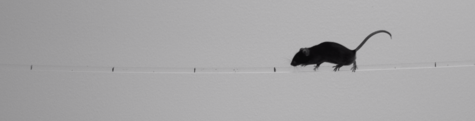
Animals without CSF-contacting neurons were slower and slipped more often when they needed to elongate and bend their body axis while balancing.
The changes only became apparent after the researchers coaxed the mice to cross a narrow balance beam or horizontal ladders, on which the animals had to elongate and bend their body axis while balancing. “The mice in which we had eliminated CSF-contacting neurons were slower and slipped more often,” reports Gerstmann. The team observed the same results when they prevented formation of the cilium of the neurons. “The cilium thus seems to be crucial for the function of the CSF-contacting neurons,” says Gerstmann. Fishes need an ion channel called PKD2L1 to control such movements. Surprisingly, this channel seems to be dispensable in this regard in mammalian cells.
“However, we don’t yet know exactly which sensory stimuli the CSF-contacting neurons register and which receptors they use for this purpose,” explains Zampieri. The team now wants to answer these questions in further experiments with the help of molecular studies, he says, adding that it is conceivable that the cells detect the flow of the cerebrospinal fluid and its chemical composition – the latter of which changes during the animals’ development, over the course of the day or when they get sick or tired.
Contact with many organs

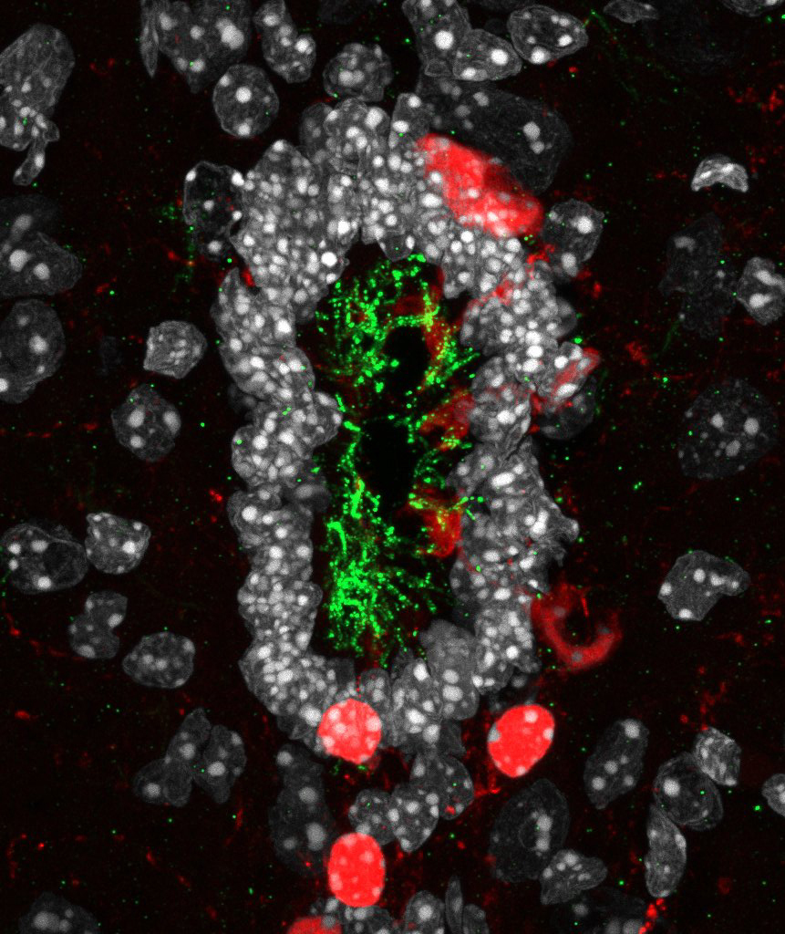
In an image at 64× magnification, the cilia (green) – antenna-like projections of the CSF-contacting neurons (red) – can also be seen.
“We also saw that the CSF-contacting neurons were not only associated with motor neurons, but also with other spinal neurons that can regulate the function of internal organs, smooth muscle cells, and gland secretion,” reports Zampieri. “The cells could therefore be part of a neuroendocrine system that also modulates other aspects of mammalian physiology.” An exciting question that remains to be answered is how the CSF-contacting neurons work with other neuronal networks to orchestrate the precise execution of complex movement patterns, the researcher says. So his sights are set on unlocking many more secrets of the spinal cord sensory neurons.
Text: Anke Brodmerkel
Further information

Literature
Katrin Gerstmann et al. (2022): “The role of intraspinal sensory neurons in the control of quadrupedal locomotion.” Current Biology, DOI: 10.1016/j.cub.2022.04.019
Downloads
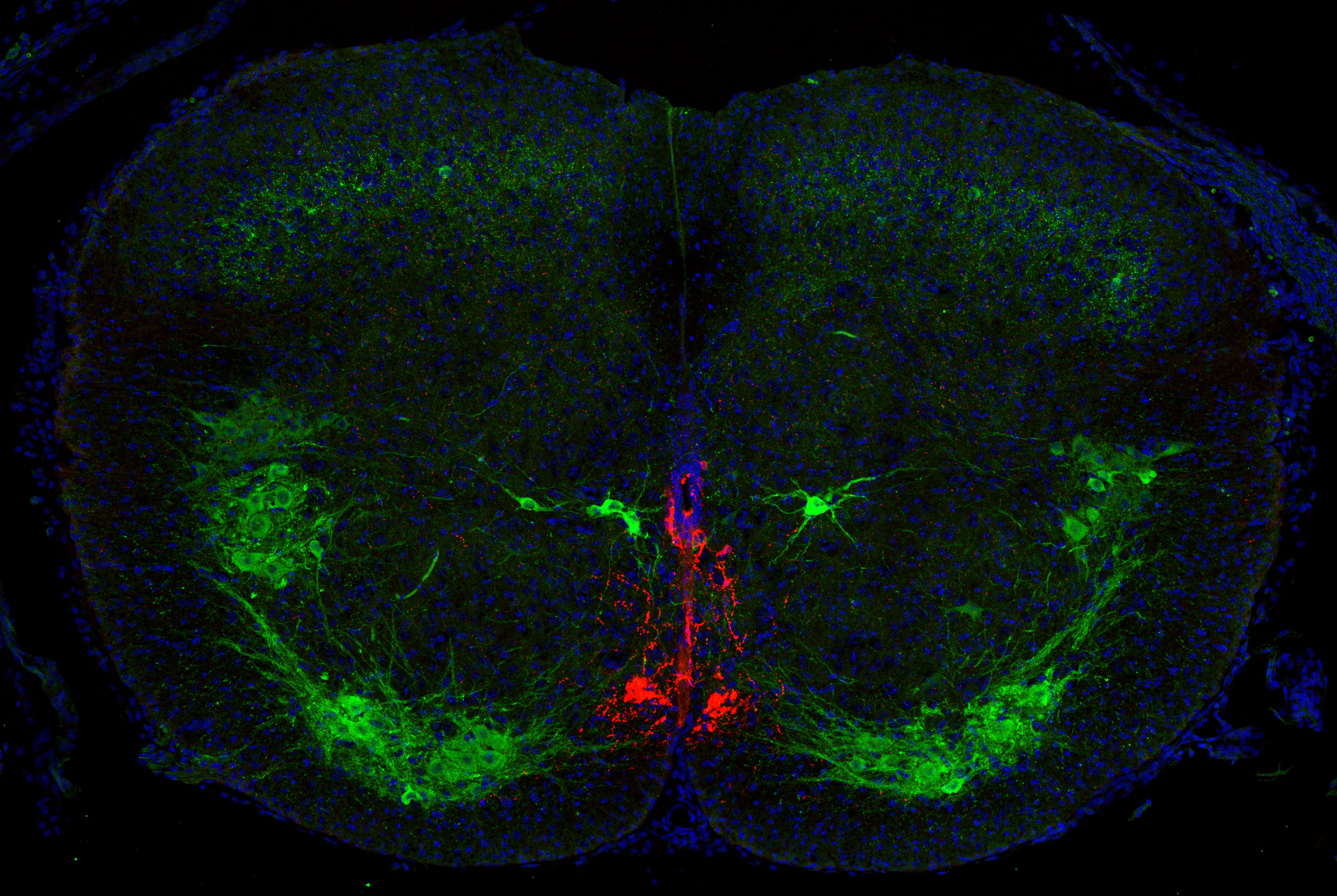
- A cross-section of the spinal central canal of a mouse. CSF-contacting cells are red, while motor neurons and another cell population near the central canal are stained green. Photo: Zampieri Lab, MDC
- In an image at 64× magnification, the cilia (green) – antenna-like projections of the CSF-contacting neurons (red) – can also be seen. Photo: Zampieri Lab, MDC
- Animals without CSF-contacting neurons were slower and slipped more often when they needed to elongate and bend their body axis while balancing. Photo: Zampieri Lab, MDC
Contacts
Dr. Niccolò Zampieri
Head of the Development and Function of Neural Circuits Lab
Max Delbrück Center for Molecular Medicine in the Helmholtz Association (MDC)
Niccolo.Zampieri@mdc-berlin.de
Jana Schlütter
Editor, Communications Department
Max Delbrück Center for Molecular Medicine in the Helmholtz Association (MDC)
+49-(0)30 – 9406-2121
jana.schluetter@mdc-berlin.de or presse@mdc-berlin.de
- Max Delbrück Center for Molecular Medicine in the Helmholtz Association (MDC)
-
The Max Delbrück Center for Molecular Medicine in the Helmholtz Association (MDC) is one of the world’s leading biomedical research institutions. Max Delbrück, a Berlin native, was a Nobel laureate and one of the founders of molecular biology. At the MDC’s locations in Berlin-Buch and Mitte, researchers from some 60 countries analyze the human system – investigating the biological foundations of life from its most elementary building blocks to systems-wide mechanisms. By understanding what regulates or disrupts the dynamic equilibrium in a cell, an organ, or the entire body, we can prevent diseases, diagnose them earlier, and stop their progression with tailored therapies. Patients should benefit as soon as possible from basic research discoveries. The MDC therefore supports spin-off creation and participates in collaborative networks. It works in close partnership with Charité – Universitätsmedizin Berlin in the jointly run Experimental and Clinical Research Center (ECRC), the Berlin Institute of Health (BIH) at Charité, and the German Center for Cardiovascular Research (DZHK). Founded in 1992, the MDC today employs 1,600 people and is funded 90 percent by the German federal government and 10 percent by the State of Berlin.


