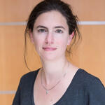
The image analyst
Certain problems make Dr. Dagmar Kainmüller’s eyes light up. When the task is to match hundreds of cells across different microscopic images or to automatically mark individual cells on an image, even huge data centers quickly reach the limits of their abilities. This is where artificial intelligence and neuronal networks come in – and where computer scientist Kainmüller is well and truly in her element. “I work at the interface of state-of-the-art biomedical research and the latest developments in automated image analysis,” says Kainmüller, who heads up the Biomedical Image Analysis Lab at the Max Delbrück Center for Molecular Medicine in the Helmholtz Association (MDC). And the bigger the mathematical challenge, the more enthusiastic she becomes.
The volume of data being produced in medicine and research is growing every year. Medical imaging alone generates petabytes of data annually. A petabyte is one quadrillion bytes (1 followed by 15 zeros). Then there are the image-based processes used in basic research, such as observing living cells or metabolic processes with microscopes. This also produces huge numbers of images that all need to be analyzed. Artificial intelligence can help the researchers and physicians do this. As well as allowing them to evaluate the images faster and more accurately, software-based analysis can also capture a wealth of additional information that’s hidden in the images.
The power of machine sight
Kainmüller’s research field is called “computer vision.” Applications for machines that can “see” range from robotics to autonomous vehicles to quality control in manufacturing. “We’re now working on making these methods fit for biomedical image analysis,” says Kainmüller, explaining the aim of her lab’s research.
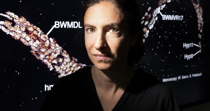
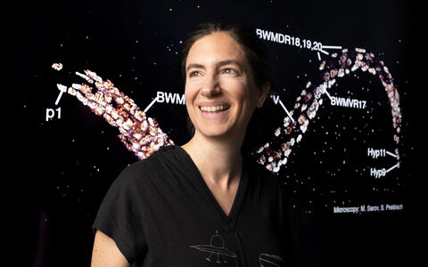
But even a small worm can present Kainmüller and her team with some big problems: The nematode C. elegans is a popular model organism in developmental biology. Juvenile worms contain exactly 558 cells that develop in exactly the same way in every worm. Cell biologists compare microscopic images of different worms and try to match the individual cells as accurately as possible. It’s a difficult and time-consuming task. “Current computing capacities aren’t enough to try all the combinatorial possibilities across two images in a finite amount of time,” says Kainmüller.
Combinatorial optimization rests on the most beautiful mathematics.
What’s needed is software that uses specific criteria to limit the combinatorial possibilities and then deliver a solution that’s as close to optimal as possible. To develop these kinds of programs, Kainmüller is working in a global network. “We publish our questions as benchmarks and encourage the international research community to publish their approaches to them.” Friendly competition with other programmers invariably leads to better solutions that ultimately benefit researchers working in computer science, artificial intelligence, and cell biology. As well as formal skills, there’s always a need for intuition, too. It‘s vital for coming up with creative new methods, says Kainmüller, who adores the mathematical puzzles that arise in her work: “Combinatorial optimization rests on the most beautiful mathematics.”
Kainmüller discovered her passion for math and computers at school in Bensheim, a town in southwest Germany. “Our computer science lessons were fantastic,” she says. This led her to study computer science with a minor in mathematics at university in Karlsruhe and Lübeck, and the Zuse Institute in Berlin. Very few women study these subjects, which Kainmüller sees as the manifestation of a glaring fault in society. Women who choose degrees in math and science still aren’t treated equally to their male counterparts: “People kept asking me how I ended up in computer science as a woman,” she says. But for her, it’s motivation: “I really want to make things better for women in IT careers.” To do so, she participates in Girls’ Day and other projects aimed at getting more women into math and science, and focuses on diversity when recruiting for her lab.
Images of nematodes, zebrafish, and axolotls
I like it when my work produces a visible outcome.
Kainmüller’s own career has been shaped by her passion for images and image analysis. On the hunt for meaningful applications for computer vision, she devoted her doctorate to image data from clinical research and spent her postdoc focusing on basic biological research. She has developed methods for analyzing microscopic images of nematodes, zebrafish, and axolotls. She sees computer science as applied mathematics, and this aspect is important to her: “I like it when my work produces a visible outcome,” she says. “A lot of computer scientists work on abstract problems that don’t have much connection to the real world.”
Kainmüller began getting involved in clinical research projects a long time ago. Right now, she’s working with Professor Andreas Hocke at the Department of Infectious Diseases and Respiratory Medicine at Charité – Universitätsmedizin Berlin. Hocke is researching how viral and bacterial infections develop at the cellular-molecular level, with a special focus on tissue left over from lung tumor surgery. This tissue can be used to make organoids, which are miniature artificial organs. They’re very similar to the human lung and allow researchers to carry out long-term studies tailored to individual patients. The team uses high-resolution fluorescence microscopy to observe how an infection spreads throughout the tissue. This produces many images that, without artificial intelligence, have to be painstakingly evaluated by hand. Kainmüller is working on methods that will be able to automatically mark individual cells and nuclei on the images. Researchers will then be able to do this type of work much faster.
Kainmüller is also working with Professor Christine Sers at the Institute of Pathology at Charité. As part of CompCancer, a PhD program funded by the German Research Foundation, they are exploring ways of extracting new information from histological images of bowel cancer using artificial intelligence. One of the main questions is whether the image data can be used to work out the likelihood of a bowel tumor metastasizing. Kainmüller explains that the artificial intelligence works like a black box: “We don’t know exactly where the AI draws the information from, but our initial results indicate that the predictions are better than random distribution.” The findings could then be channeled back into medical research as a new question: Of the cell biological properties discernible in the images, which ones affect the development of metastases?
More mathematical puzzles waiting in the wings
The computer scientists are also using the tiny slices of tissue to try and work out how best to treat an individual tumor. Their prediction models will also include data from gene sequences of neighboring cells and from trials of various therapeutic agents in individually cultivated organoids. “In future, we might be able to use the microscopic image of a piece of tissue to predict the best therapy for individual patients,” says Kainmüller.
Her long-term goal is to develop a model for computer-aided image analysis that can be used for multiple applications. Kainmüller and MDC structural biologist Professor Oliver Daumke have recently received funding from Helmholtz Imaging for their work.
Berlin is full of new research questions and fields of application.
“New imaging techniques are constantly being developed, but there are no data available for training the artificial intelligence,” says Kainmüller, explaining the challenge. In future, artificial intelligence should be able to use independent machine learning to identify the important aspects of the image data in question. But there are still quite a few mathematical puzzles to solve before it’s ready for use. “Berlin is full of new research questions and fields of application,“ says Kainmüller. “It will be my playground for a long time yet.”
Text: Dietrich von Richthofen

