
The decrypter of cryptic proteins
On a gray winter day, Dr. Melissa Birol sits at a round table in her office, sketching on a whiteboard next to her. She draws cells, their organelles and strange squiggly lines around them. Birol is Group Leader of the Single Molecule Biophysics probing Quantitative Neuroscience lab at the Berlin Institute for Medical Systems Biology of the Max Delbrück Center (MDC-BIMSB). With her minimalist illustrations, Birol explains how she is developing new models to study unusual proteins that lack a stable 3D structure – or intrinsically disordered proteins (IDPs) – and how they ultimately lead to devastating neurological diseases such as Alzheimer’s and Parkinson’s.

Up to 40% of proteins in our bodies are disordered. For years, researchers neglected to study them because they weren’t considered important, says Birol.
We now know that IDPs are involved in many diseases including cancer, diabetes, cardiovascular and incurable neurological conditions such as Alzheimer’s, Parkinson’s and Amyotrophic Lateral Sclerosis. In brain diseases, they form the basic components of the characteristic clumps, known as pathological plaques, found in the brain and nervous system tissue of patients. And yet, no one knows what their normal functions in the body are, or how they accumulate.
Armed with new organoid models that more closely resemble the human brain and sophisticated imaging technology, Birol aims to better understand the normal function of these curious proteins and how they go rogue, eventually forming tangled clusters in the brain.
From physics to biomedicine
Birol wasn’t always interested in biology. During her high school years in Greece, she focused on physics, math, chemistry and art. As an undergraduate, she considered majoring in design because she liked to paint and create things. But her parents, who are both chemical engineers, vetoed the idea. “They told me I could always paint,” she recalls. She followed their advice – and not necessarily reluctantly. Physics always fascinated her, she says, so she decided to major in the subject.
During a Master’s program at Imperial College in London, she used theoretical physics and structural modelling to understand how the Rubisco protein in plants captures CO2 from the environment – it was her first foray into the biological sciences.
While earning her doctorate at the Center for Structural Biology in Montpellier, France, Birol was using 3D x‑ray crystallography images and computer simulation models to understand the dynamic structure of a protein called COP9, which helps degrade malfunctioning proteins in cells. But x‑ray crystallography only takes snapshots of a molecule at a single point in time. Meanwhile, some of the regions of the enzymatic subunit of the COP9 protein complex are disordered – they move and change shape, forming loops at times.
“What intrigued me was that there were always regions we couldn’t model. They were so dynamic that they don’t exist in the crystal structure,” Birol says. “I became really interested in IDPs and finding tools that would allow us to understand their function better.”
Shape-shifters come of age
Most proteins fold into specific 3D structures – shapes that are encoded in our genes. Throughout their lifecycle they remain folded and carry out specific tasks. But some proteins are promiscuous. After they are translated by the cell’s protein-making machinery, they change shapes depending on the environment they encounter, almost like strands of spaghetti.
For most of the last century, structural biology had been dominated by the idea that a protein’s function was dictated by its stable, three-dimensional shape. Any proteins that didn’t fit this narrative were quietly ignored.
By 2010, however, it had become clear that IDPs play a major role in neurological disease. Studies of the characteristic plaques that form in the brains of people with Parkinson’s and Alzheimer’s, for example, were showing that they contained a mess of disordered proteins among other “junk,” says Birol. The proteins inside these plaques are no longer disordered, but folded, which is why these plaques are sometimes referred to as containing “misfolded proteins.”
Today, researchers still don’t understand the normal function of these proteins. Even more perplexing is why they form pathological plaques. Take alpha-synuclein, for example, a protein that makes up a central component of Lewy Body clumps. It is found in the brains of people with Parkinson’s disease, both inside and outside cells. But no one understands what it does, Birol explains.
“We don’t know how it moves from inside the cell to the outside, whether it takes on a different structure, how it functions once outside, how it spreads, and why it ends up in these plaques.” The same is true for amyloid-beta and tau, disordered proteins that form clumps in the brains of people with Alzheimer’s.
Over the past 30 years, dozens of experimental drugs to treat Alzheimer’s and Parkinson’s disease by clearing amyloid-beta or alpha-synuclein clumps from the brain have failed in clinical trials. The few drugs that are available have only modest effect in relieving symptoms – none offer a cure. “We really need novel ways to address these diseases,” says Birol.
Studying single molecules
At the Max Delbrück Center, Birol’s team is developing new models to understand the function of alpha-synuclein, amyloid beta, tau and other disordered proteins implicated in neurological diseases.
One project aims to develop a brain organoid model of Alzheimer’s disease that integrates optogenetics – a technique that uses light to control molecules in living cells. By fusing light-sensitive proteins to amyloid-beta and exposing them to blue light, Birol can trigger amyloid-beta to spread. In this way, she aims to study the events before plaques actually form, step-by-step.
Birol can also grow organoids that contain a mix of brain cells such as neurons and glial cells and study how these cells communicate with each other, making them a good model of what is actually happening in human brains, she says. And she aims to engineer organoids to study communication between the forebrain and midbrain for example, to observe how disordered proteins move between brain regions, which will hopefully yield insight into the very early stages of neurological disease, she says.
“We think that spread of IDPs impacts the communication between different brain cell types,” Birol explains. “Treatments that target these early events might thus be more effective in preventing clumps.”
She also wants to learn whether disordered proteins such as amyloid-beta and alpha synuclein interact with each other, if at all, because neurological diseases share common pathologies. About half of Alzheimer’s patients, for example, have alpha-synuclein clumps in addition to tau and amyloid-beta in their brains, and no one knows why, she says.
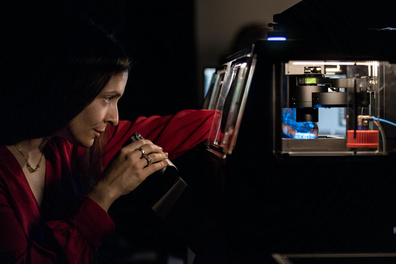
Imaging the abstract
Back in her office, the strange squiggly lines Birol is drawing represent alpha- synuclein. As the protein spreads, it is often found sitting on the flanks of mitochondria and endoplasmic reticulum – cellular organelles that communicate to exchange material in cells. Her lab is using advanced imaging approaches like fluorescence lifetime imaging microscopy (FLIM) to understand how and where the protein moves.

She and her team are also applying and developing single-molecule spectroscopy and functional metabolic and calcium imaging to their research – technologies that enable them to capture changes – from the nano to the meso to the macroscale – in individual cells over time. Such imaging can provide clues about how the shifting metabolism of cells affects such cellular characteristics as the distribution of lipids. Because lipids become concentrated inside the brain plaques of Alzheimer’s and Parkinson’s patients, such changes in brain lipid distribution as we age may fuel the spread of IDPs and neurological disease.
For Birol, tinkering with microscopes is more than a job – it has become a passion. Whether her fascination with imaging is related to her penchant for art and design, Birol isn’t sure. But she is apparently very good at it. Her former advisor from Philadelphia uses Birol’s stunning microscopy images as examples of the level of detail her students should strive to attain, she says with a grin. “Imaging, specifically multiscale imaging, is a great tool. It helps to make the abstract and complex tangible. It doesn’t answer all my questions, but our approach of systems-level imaging promises to give a more holistic understanding of these complex disease landscapes.”
Text: Gunjan Sinha


