We are an international, highly interdisciplinary group of scientists — some of us “hard-core” data scientists, others “hard-core” molecular biologists, etc — learning from each other and supporting each other. We enjoy working in a modern lab environment (data science/analyses only separated by sliding glass doors from the wet lab) in a highly collaborative institute in the heart of Berlin. We also collaborate with many colleagues across the globe (e.g. Rajewsky et al Nature 2020). We have strong ties with the nearby Charité, Europe’s largest Hospital.
Research interests and approaches
How do our genes interact to create the bewildering diversity of cell types, tissues, and organs? Can we identify perturbations of these interactions that cause or drive the onset of diseases? And how can we predict and target early disease trajectory for therapy before symptoms appear?
To answer these questions, we pursue, for 20 years, a strongly interdisciplinary approach by integrating biochemistry, molecular biology, and data sciences. We are mainly working with patient samples (numerous collaboration with the Charité hospital Berlin) and patient-derived organoids. Our research questions often require us to develop or apply new methods to quantify how cells read and interpret the book of life (the genome) in health and in disease. We use data sciences (including machine learning) and our brains to interpret these data and to make predictions about mechanisms which we then test directly in the lab. A long-standing special interest of the lab is to understand the function of the non-coding genome/RNA (miRNAs, circRNAs, long-non-coding RNAs, …).
Diseases currently studied in the lab: Cancer (Triple negative breast cancer, non-small cell lung cancer, neuroblastoma), Neurodegenerative diseases and impact of infections on brain development.
Lieke van de Haar was a Postdoctoral Researcher. Current: patent lawyer
Enes Senel was a PhD Student. Current: Senior Scient at J&J Innovative Medicine.
Seung Kim was a PhD Student. Current: Scientist at EBIUK
Tamas Ryszard Sztanka-Toth was a PhD Student. Current: Principal Scient at J&J Innovative Medicine.
Haiyue Liu was a PhD Student
Agnieszka Rybak-Wolf was a Postdoctoral Researcher. Current: Independent Head of the MDC Technology Platform “Organoids”.
Jonathan Alles was a PhD Student. Current: Scientist, Evobyte Digital Biology
Ivano Legnini was a Postdoctoral Researcher. Current: Group Leader, Human Technopole, Mailand, Italy.
Petar Glažar was a PhD Student and Postdoctoral Researcher. Current: Postdoc at MPI Molecular Genetics, Aktas lab.
Jonathan Fröhlich was a PhD Student.
Aristotelis Misios was a PhD Student.
Samantha Praktiknjo was a Postdoctoral Researcher. Current: Scientist at Berlin Institute for Health.
Marcel Schilling was a PhD Student.
Janis Hötzel was a PhD Student.
Monika Piwecka was a Postdoctoral Researcher. Current: Independent Group Leader at Institute of Bioorganic Chemistry, Polish Academy of Science, Poznan.
Mireya Plass Pórtulas was a Postdoctoral Researcher. Current: Independent Group Leader at Institut d’Investigació Biomédica de Bellvitge (IDIBELL) and Clinical Translation of Regenerative Medicine of Catalonia (P‑CMR[C]), Barcelona, Spain.
Christin Sünkel (née Stottmeister) was a PhD Student. Current: Technical Project Analyst, Lonza, Basel.
Kathrin Theil was a PhD Student. Current: Scientist at Labor Berlin – Charité Vivantes.
Panagiotis Papavasileiou was a PhD Student.
Christine Kocks was a Senior Scientist.
Vera Zywitza was a PhD Student. Current: Scientist at the Leibniz Institute for Zoo and Wildlife Research.
Sebastian Memczak was a PhD Student and Postdoctoral Researcher. Current: Postdoc at Salk Institute, Belmonte lab.
Benedikt Obermayer was a Postdoctoral Researcher. Current: Bioinformatician at BIH Core Unit Bioinformatics, Berlin.
Andrei Filipchyk was a PhD Student. Current: Application Expert, Evotec.
Marta Rodriguez-Orejuela was a PhD Student.
Luisa Schreyer was a Technical Assistant.
Jordi Solana Garcia was a Postdoctoral Researcher. Current: Independent Group Leader at Oxford Brookes University, Oxford, UK.
Filippos Klironomos was a Postdoctoral Researcher. Current: Bioinformatics Data Scientists, Nuvisan Pharma services.
Catherine Adamidi (†) was a Senior Scientist.
Kevin Chen was a Postdoctoral Researcher. Current: Professor at Rutgers University.
Antigoni Elefsinioti was a Postdoctoral Researcher. Current: Bayer
Marc Friedländer was a PhD Student and Postdoctoral Researcher. Current: Associate Professor at Stockholm University.
Stefanie Grosswendt was a PhD Student. Current: Lise-Meitner-Fellow at Max-Planck-Gesellschaft.
Dominic Grün was a Postdoctoral Researcher. Current: Professor at University Würzburg.
Andranik Ivanov was a PhD Student. Current: Independent Group Leader, Human Technopole, Mailand, Italy.
Anna-Carina Jungkamp was a PhD Student. Current: Federal Ministry of Research, Technology and Space in Berlin, Germany.
Toshiaki Kogame was a PhD Student.
Azra Krek was a PhD Student.
Svetlana Lebedeva was a PhD Student.
Jonas Maaskola was a PhD Student.
Sebastian Mackowiak was a PhD Student.
Pinar Önal was a PhD Student. Current: Professor, BILKENT University, Ankara, Turkey.
Lena von Oertzen was a Technical Assistant.
Marlon Stoeckius was a PhD Student and Postdoctoral Researcher.
Nadine Thierfelder was a PhD Student. Current: Medical Advisor at Besins Healthcare Germany.
Francesca Torti was a PhD Student.
Ulrike Ziebold was a Postdoctoral Researcher.
Minnie Zhou Fang was a Postdoctoral Researcher. Current: CTO at Wuxi Diagnostics, Shanghai, China.
Research
The complete list of publications is available on Google Scholar.
Recently, we developed an inexpensive, do-it-yourself, high-resolution, and open-source method to quantify gene expression directly in tissues (Schott, Leon-Perinān, Splendiani et al. “Open-ST: High-resolution spatial transcriptomics in 3D” 2024, Cell; 2024, STAR Protocols; Github).
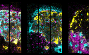
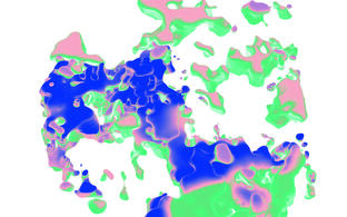
We constructed the first high-resolution molecular tumor atlas in 3D (from a single patient) and learned from these ~1 million sequenced cells (in tissue space) which pathways and gene programs drive phenotypes of the primary and metastatic tumor.
We currently routinely generate spatial transcriptomics data as a highly informative readout for how cells communicate within tissues/tumors and how they react to perturbations (disease mutations, perturbations from the environment, viral infection, microplastics, etc).
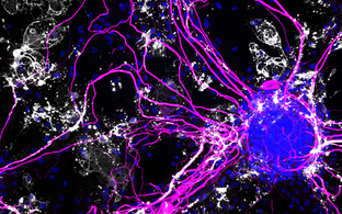
Organoids
In a human brain organoid model, we found that Herpes induced neural damage after Herpes activation (a debilitating clinical problem and practically impossible to study in patients) can be prevented in brain organoids by treating them not only with acyclovir (the standard drug given in the clinics) but also with inflammation blockers Rybak-Wolf et al., 2023, Nature Microbiology.
We also invented an optogenetic approach to perturb expression of specific genes at precisely located regions/cells within organoids Legnini et al., 2023, Nature Methods. This approach allows, for example, to work with “programmed organoids” to test specific questions about how gene interactions organize tissue function in health and disease.
We could also show that miRNA evolution in vertebrates and the octopus share common global features (Zolotarov et al., 2022, Science Advances) and, excited by these insights, are now studying the function of primate specific miRNAs in human brain development.
Whether your weapon of choice is pipette or keyboard (or both): if you have the impression that this lab is doing interesting research that you would like to contribute to, do not hesitate to contact us.
If you are able to (and enjoy) discussing your work with people from other disciplines this lab may be just right!
We are located within a few minutes walk from several public transport stations, such as Oranienburger Tor, Oranienburger Straße, and only one stop away from the public transport hub Friedrichstraße. From Oranienburger Straße, the train S2 connects us directly to the MDC Campus in Berlin-Buch.


