ATTRACT: Arterial flow as attractor for endothelial cell migration
Fondation Leducq Transatlantic Network of Excellence
The project
What is Leducq ATTRACT about?
The cells that form the inner lining of arteries are called endothelial cells. It was once thought that endothelial cells were an immobile interface between the blood flowing past them and the tissues which are perfused with oxygen. However, recent discoveries by our network’s members have shown that endothelial cells can change position within vessels, and that they actually have been observed to migrate in the opposite direction of blood flow. This finding has led us to examine whether this endothelial cell migration plays a role in the normal development of blood vessels. In addition, we hope to understand how endothelial cell migration may play a role in disease. In vascular disease such as stroke and coronary artery disease, the atherosclerotic process disrupts the endothelial layer and changes the pattern of cell migration. Our network will investigate the principles of normal vessel function and how endothelial cell movement and repair are maladaptive after a stroke. We hope to learn how to harness the beneficial movement of the endothelial cells so that patients with neurovascular disease have better outcomes, and abnormal vascular connections are avoided.
The Integrative Vascular Biology Laboratory aims to unravel the fundamental principles and molecular/genetic regulation of functional vascular network formation in development and disease.
The Vascular Morphogenesis Lab has the aim to understand the molecular mechanisms regulating coordinated endothelial cell behaviour during sprouting and remodelling phases of the angiogenic process.
Lena’s lab studies how blood vessels are formed and which factors control the process, positively or negatively. Major topics of her research are how vascular endothelial growth factor (VEGF) regulates angiogenesis and the signal transduction pathways which regulate blood vessel permeability.
Miguel is a Computational Biologist specialised in microvascular Biomechanics and Mechanobiology. His research interests concern the study of vascular remodelling during angiogenesis in order to identify molecular targets for its regulation. In addition, he explores the translational potential of these findings for the treatment diabetic retinopathy.
Paul Oh’s research interests include identifying cellular mechanisms responsible for cerebrovascular disorders and assessing novel therapies to treat these disorders.
The Molecular Stroke Research Lab investigates vascular and neuronal network plasticity in rodent stroke models. They aim to explore genetic/inflammatory/imaging surrogates that predict outcome and identify interventions to retune disabled vascular and neuronal networks with a particular focus on translation.
Publications
Here you can find the most recent publications from the network groups, relevant for the Leducq project.
VEGFR2 Expression Correlates with Postnatal Development of Brain Arteriovenous Malformations in a Mouse Model of Type I Hereditary Hemorrhagic Telangiectasia
November 2023,Biomedcines.Chul Han,Candice L Nguyen, Lea Scherschinski, Tyler D Schriber, Helen M Arthur, Michael T Lawton, and S. Paul Oh.
Abstract
Brain arteriovenous malformations (BAVMs) are a critical concern in hereditary hemorrhagic telangiectasia (HHT) patients, carrying the risk of life-threatening intracranial hemorrhage. While traditionally seen as congenital, the debate continues due to documented de novo cases. Our primary goal was to identify the precise postnatal window in which deletion of the HHT gene Endoglin (Eng) triggers BAVM development. We employed SclCreER(+);Eng2f/2f mice, enabling timed Eng gene deletion in endothelial cells via tamoxifen. Tamoxifen was given during four postnatal periods: P1‑3, P8-10, P15-17, and P22-24. BAVM development was assessed at 2 – 3 months using latex dye perfusion. We examined the angiogenic activity by assessing vascular endothelial growth factor receptor 2 (VEGFR2) expression via Western blotting and Flk1-LacZ reporter mice. Longitudinal magnetic resonance angiography (MRA) was conducted up to 9 months. BAVMs emerged in 88% (P1‑3), 86% (P8-10), and 55% (P15-17) of cases, with varying localization. Notably, the P22-24 group did not develop BAVMs but exhibited skin AVMs. VEGFR2 expression peaked in the initial 2 postnatal weeks, coinciding with BAVM onset. These findings support the “second hit” theory, highlighting the role of early postnatal angiogenesis in initiating BAVM development in HHT type I mice.
Single-cell profiling and zebrafish avatars reveal LGALS1 as immunomodulating target in glioblastoma
November 2023,EMBO Mol Med.Lise Finotto, Basiel Cole, Wolfgang Giese, Elisabeth Baumann, Annelies Claeys, Maxime Vanmechelen, Brecht Decraene, Marleen Derweduwe, Nikolina Dubroja Lakic, Gautam Shankar, Madhu Nagathihalli Kantharaju, Jan Philipp Albrecht, Ilse Geudens, Fabio Stanchi, Keith L Ligon, Bram Boeckx, Diether Lambrechts, Kyle Harrington, Ludo Van Den Bosch, Steven De Vleeschouwer, Frederik De Smet, Holger Gerhardt
Abstract
Glioblastoma (GBM) remains the most malignant primary brain tumor, with a median survival rarely exceeding 2 years. Tumor heterogeneity and an immunosuppressive microenvironment are key factors contributing to the poor response rates of current therapeutic approaches. GBM-associated macrophages (GAMs) often exhibit immunosuppressive features that promote tumor progression. However, their dynamic interactions with GBM tumor cells remain poorly understood. Here, we used patient-derived GBM stem cell cultures and combined single-cell RNA sequencing of GAM-GBM co-cultures and real-time in vivo monitoring of GAM-GBM interactions in orthotopic zebrafish xenograft models to provide insight into the cellular, molecular, and spatial heterogeneity. Our analyses revealed substantial heterogeneity across GBM patients in GBM-induced GAM polarization and the ability to attract and activate GAMs-features that correlated with patient survival. Differential gene expression analysis, immunohistochemistry on original tumor samples, and knock-out experiments in zebrafish subsequently identified LGALS1 as a primary regulator of immunosuppression. Overall, our work highlights that GAM-GBM interactions can be studied in a clinically relevant way using co-cultures and avatar models, while offering new opportunities to identify promising immune-modulating targets.
Prediction of Stroke Outcome in Mice Based on Noninvasive MRI and Behavioral Testing
September 2023, Stroke Felix Knab, Stefan Paul Koch, Sebastian Major, Tracy D Farr, Susanne Mueller, Philipp Euskirchen, Moritz Eggers, Melanie T C Kuffner, Josefine Walter, Daniel Berchtold, Samuel Knauss, Jens P Dreier, Andreas Meisel, Matthias Endres, Ulrich Dirnagl, Nikolaus Wenger, Christian J Hoffmann, Philipp Boehm-Sturm, Christoph Harms
Abstract
Background: Prediction of poststroke outcome using the degree of subacute deficit or magnetic resonance imaging is well studied in humans. While mice are the most commonly used animals in preclinical stroke research, systematic analysis of outcome predictors is lacking.
Methods: We intended to incorporate heterogeneity into our retrospective study to broaden the applicability of our findings and prediction tools. We therefore analyzed the effect of 30, 45, and 60 minutes of arterial occlusion on the variance of stroke volumes. Next, we built a heterogeneous cohort of 215 mice using data from 15 studies that included 45 minutes of middle cerebral artery occlusion and various genotypes. Motor function was measured using a modified protocol for the staircase test of skilled reaching. Phases of subacute and residual deficit were defined. Magnetic resonance images of stroke lesions were coregistered on the Allen Mouse Brain Atlas to characterize stroke topology. Different random forest prediction models that either used motor-functional deficit or imaging parameters were generated for the subacute and residual deficits.
Results: Variance of stroke volumes was increased by 45 minutes of arterial occlusion compared with 60 minutes. The inclusion of various genotypes enhanced heterogeneity further. We detected both a subacute and residual motor-functional deficit after stroke in mice and different recovery trajectories could be observed. In mice with small cortical lesions, lesion volume was the best predictor of the subacute deficit. The residual deficit could be predicted most accurately by the degree of the subacute deficit. When using imaging parameters for the prediction of the residual deficit, including information about the lesion topology increased prediction accuracy. A subset of anatomic regions within the ischemic lesion had particular impact on the prediction of long-term outcomes. Prediction accuracy depended on the degree of functional impairment.
Conclusions: For the first time, we developed and validated a robust tool for the prediction of functional outcomes after experimental stroke in mice using a large and genetically heterogeneous cohort. These results are discussed in light of study design and imaging limitations. In the future, using outcome prediction can improve the design of preclinical studies and guide intervention decisions.
Keywords: forecasting; infarction; mice, Inbred C57BL; middle cerebral artery; precision medicine; random forest; sensory motor performance; topographic brain mapping.
A 96-wells fluidic system for high-throughput screenings under laminar high wall shear stress conditions
September 2023, Microsyst Nanoeng.Catarina Gonçalves Fonseca, Vânia Silvério, David Barata, Wolfgang Giese, Holger Gerhardt, Susana Cardoso, Claudio Areias Franco
Abstract
The ability of endothelial cells to respond to blood flow is fundamental for the correct formation and maintenance of a functional and hierarchically organized vascular network. Defective flow responses, in particular related to high flow conditions, have been associated with atherosclerosis, stroke, arteriovenous malformations, and neurodegenerative diseases. Yet, the molecular mechanisms involved in high flow response are still poorly understood. Here, we described the development and validation of a 96-wells fluidic system, with interchangeable cell culture and fluidics, to perform high-throughput screenings under laminar high-flow conditions. We demonstrated that endothelial cells in our newly developed 96-wells fluidic system respond to fluid flow-induced shear stress by aligning along the flow direction and increasing the levels of KLF2 and KLF4. We further demonstrate that our 96-wells fluidic system allows for efficient gene knock-down compatible with automated liquid handling for high-throughput screening platforms. Overall, we propose that this modular 96-well fluidic system is an excellent platform to perform genome-wide and/or drug screenings to identify the molecular mechanisms involved in the responses of endothelial cells to high wall shear stress.
Endothelial VEGFR2-PLCγ signaling regulates vascular permeability and anti-tumor immunity through eNOS/Src
August 2023, J Clin InvestElin Sjöberg, Marit Melssen, Mark Richards, Yindi Ding, Catarina Chanoca, Dongying Chen, Emmanuel Nwadozi, Sagnik Pal, Dominic T Love, Takeshi Ninchoji, Masabumi Shibuya, Michael Simons, Anna Dimberg, Lena Claesson-Welsh
Abstract
Endothelial phospholipase Cγ (PLCγ) is essential for vascular development, however, its role in healthy, mature or pathological vessels is unexplored. PLCγ was prominently expressed in vessels of several human cancer forms, notably in renal cell carcinoma (RCC). High PLCγ expression in clear cell RCC correlated with angiogenic activity and poor prognosis, while low expression correlated with immune cell activation. PLCγ was induced downstream of vascular endothelial growth factor receptor 2 (VEGFR2) phosphosite Y1173 (pY1173). Heterozygous Vegfr2+/Y1173F mice or mice lacking endothelial PLCγ (Plcg1iECKO) exhibited a stabilized endothelial barrier and diminished vascular leakage. Barrier stabilization was accompanied by decreased expression of immunosuppressive cytokines, reduced infiltration of B‑cells, CD4+ and regulatory T‑cells, and improved response to chemo- and immunotherapy. Mechanistically, pY1173/PLCγ signaling induced Ca2+/protein kinase C dependent activation of endothelial nitric oxide synthase (eNOS), required for tyrosine nitration and activation of Src. Src-induced phosphorylation of VE-cadherin at Y685 was accompanied by disintegration of endothelial junctions. This pY1173/PLCγ/eNOS/Src pathway was detected in both healthy and tumor vessels in Vegfr2Y1173F/+ mice, which displayed decreased activation of PLCγ and eNOS, and suppressed vascular leakage. Thus, we have identified a clinically relevant endothelial PLCγ pathway downstream of VEGFR2 pY1173, which destabilizes the endothelial barrier resulting in loss of anti-tumor immunity.
Keywords: Oncology; Signal transduction; Vascular Biology.
Chylomicrons Regulate Lacteal Permeability and Intestinal Lipid Absorption
August 2023, Circ ResGeorgia Zarkada, Xun Chen, Xuetong Zhou, Martin Lange, Lei Zeng, Wenyu Lv, Xuan Zhang, Yunhua Li, Weibin Zhou, Keli Liu, Dongying Chen, Nicolas Ricard, James Liao, Young-Bum Kim, Rui Benedito, Lena Claesson-Welsh, Kari Alitalo, Michael Simons, Rong Ju, Xuri Li, Anne Eichmann, Feng Zhang
Background: Lymphatic vessels are responsible for tissue drainage, and their malfunction is associated with chronic diseases. Lymph uptake occurs via specialized open cell-cell junctions between capillary lymphatic endothelial cells (LECs), whereas closed junctions in collecting LECs prevent lymph leakage. LEC junctions are known to dynamically remodel in development and disease, but how lymphatic permeability is regulated remains poorly understood.
Methods: We used various genetically engineered mouse models in combination with cellular, biochemical, and molecular biology approaches to elucidate the signaling pathways regulating junction morphology and function in lymphatic capillaries.
Results: By studying the permeability of intestinal lacteal capillaries to lipoprotein particles known as chylomicrons, we show that ROCK (Rho-associated kinase)-dependent cytoskeletal contractility is a fundamental mechanism of LEC permeability regulation. We show that chylomicron-derived lipids trigger neonatal lacteal junction opening via ROCK-dependent contraction of junction-anchored stress fibers. LEC-specific ROCK deletion abolished junction opening and plasma lipid uptake. Chylomicrons additionally inhibited VEGF (vascular endothelial growth factor)-A signaling. We show that VEGF‑A antagonizes LEC junction opening via VEGFR (VEGF receptor) 2 and VEGFR3-dependent PI3K (phosphatidylinositol 3‑kinase)/AKT (protein kinase B) activation of the small GTPase RAC1 (Rac family small GTPase 1), thereby restricting RhoA (Ras homolog family member A)/ROCK-mediated cytoskeleton contraction.
Conclusions: Our results reveal that antagonistic inputs into ROCK-dependent cytoskeleton contractions regulate the interconversion of lymphatic junctions in the intestine and in other tissues, providing a tunable mechanism to control the lymphatic barrier.
Deep learning-based automated lesion segmentation on mouse stroke magnetic resonance images
August 2023, Sci Rep. Jeehye An, Leo Wendt, Georg Wiese, Tom Herold, Norman Rzepka, Susanne Mueller, Stefan Paul Koch, Christian J Hoffmann, Christoph Harms, and Philipp Boehm-Sturm
Abstract
Magnetic resonance imaging (MRI) is widely used for ischemic stroke lesion detection in mice. A challenge is that lesion segmentation often relies on manual tracing by trained experts, which is labor-intensive, time-consuming, and prone to inter- and intra-rater variability. Here, we present a fully automated ischemic stroke lesion segmentation method for mouse T2-weighted MRI data. As an end-to-end deep learning approach, the automated lesion segmentation requires very little preprocessing and works directly on the raw MRI scans. We randomly split a large dataset of 382MRI scans into a subset (n = 293) to train the automated lesion segmentation and a subset (n = 89) to evaluate its performance. We compared Dice coefficients and accuracy of lesion volume against manual segmentation, as well as its performance on an independent dataset from an open repository with different imaging characteristics. The automated lesion segmentation produced segmentation masks with a smooth, compact, and realistic appearance that are in high agreement with manual segmentation. We report dice scores higher than the agreement between two human raters reported in previous studies, highlighting the ability to remove individual human bias and standardize the process across research studies and centers.
Localized conditional induction of brain arteriovenous malformations in a mouse model of hereditary hemorrhagic telangiectasia
May 2023, AngiogenesisLea Scherschinski, Chul Han, Yong Hwan Kim, Ethan A Winkler, Joshua S Catapano, Tyler D Schriber, Peter Vajkoczy, Michael T Lawton, S Paul Oh
Background: Longitudinal mouse models of brain arteriovenous malformations (AVMs) are crucial for developing novel therapeutics and pathobiological mechanism discovery underlying brain AVM progression and rupture. The sustainability of existing mouse models is limited by ubiquitous Cre activation, which is associated with lethal hemorrhages resulting from AVM formation in visceral organs. To overcome this condition, we developed a novel experimental mouse model of hereditary hemorrhagic telangiectasia (HHT) with CreER-mediated specific, localized induction of brain AVMs.
Methods: Hydroxytamoxifen (4‑OHT) was stereotactically delivered into the striatum, parietal cortex, or cerebellum of R26CreER; Alk12f/2f (Alk1-iKO) littermates. Mice were evaluated for vascular malformations with latex dye perfusion and 3D time-of-flight magnetic resonance angiography (MRA). Immunofluorescence and Prussian blue staining were performed for vascular lesion characterization.
Results: Our model produced two types of brain vascular malformations, including nidal AVMs (88%, 38⁄43) and arteriovenous fistulas (12%, 5⁄43), with an overall frequency of 73% (43÷59). By performing stereotaxic injection of 4‑OHT targeting different brain regions, Alk1-iKO mice developed vascular malformations in the striatum (73%, 22⁄30), in the parietal cortex (76%, 13⁄17), and in the cerebellum (67%, 8⁄12). Identical application of the stereotaxic injection protocol in reporter mice confirmed localized Cre activity near the injection site. The 4‑week mortality was 3% (2÷61). Seven mice were studied longitudinally for a mean (SD; range) duration of 7.2 (3; 2.3−9.5) months and demonstrated nidal stability on sequential MRA. The brain AVMs displayed microhemorrhages and diffuse immune cell invasion.
Conclusions: We present the first HHT mouse model of brain AVMs that produces localized AVMs in the brain. The mouse lesions closely resemble the human lesions for complex nidal angioarchitecture, arteriovenous shunts, microhemorrhages, and inflammation. The model’s longitudinal robustness is a powerful discovery resource to advance our pathomechanistic understanding of brain AVMs and identify novel therapeutic targets.
Fine-mapping of retinal vascular complexity loci identifies Notch regulation as a shared mechanism with myocardial infarction outcomes
May 2023, Commun BiolAna Villaplana-Velasco, Marie Pigeyre, Justin Engelmann, Konrad Rawlik, Oriol Canela-Xandri, Claire Tochel, Frida Lona-Durazo, Muthu Rama Krishnan Mookiah, Alex Doney, Esteban J Parra, Emanuele Trucco, Tom MacGillivray, Kristiina Rannikmae, Albert Tenesa, Erola Pairo-Castineira, Miguel O Bernabeu
There is increasing evidence that the complexity of the retinal vasculature measured as fractal dimension, Df, might offer earlier insights into the progression of coronary artery disease (CAD) before traditional biomarkers can be detected. This association could be partly explained by a common genetic basis; however, the genetic component of Df is poorly understood. We present a genome-wide association study (GWAS) of 38,000 individuals with white British ancestry from the UK Biobank aimed to comprehensively study the genetic component of Df and analyse its relationship with CAD. We replicated 5 Df loci and found 4 additional loci with suggestive significance (P < 1e-05) to contribute to Df variation, which previously were reported in retinal tortuosity and complexity, hypertension, and CAD studies. Significant negative genetic correlation estimates support the inverse relationship between Df and CAD, and between Df and myocardial infarction (MI), one of CAD’s fatal outcomes. Fine-mapping of Df loci revealed Notch signalling regulatory variants supporting a shared mechanism with MI outcomes. We developed a predictive model for MI incident cases, recorded over a 10-year period following clinical and ophthalmic evaluation, combining clinical information, Df, and a CAD polygenic risk score. Internal cross-validation demonstrated a considerable improvement in the area under the curve (AUC) of our predictive model (AUC = 0.770 ± 0.001) when comparing with an established risk model, SCORE, (AUC = 0.741 ± 0.002) and extensions thereof leveraging the PRS (AUC = 0.728 ± 0.001). This evidences that Df provides risk information beyond demographic, lifestyle, and genetic risk factors. Our findings shed new light on the genetic basis of Df, unveiling a common control with MI, and highlighting the benefits of its application in individualised MI risk prediction.
Associations of clonal hematopoiesis with recurrent vascular events and death in patients with incident ischemic stroke
February 2023, BloodChristopher M Arends, Thomas G Liman, Paulina M Strzelecka, Anna Kufner, Pelle Löwe, Shufan Huo, Catarina M Stein, Sophie K Piper, Marlon Tilgner, Pia S Sperber, Savvina Dimitriou, Peter U Heuschmann, Raphael Hablesreiter, Christoph Harms, Lars Bullinger, Joachim E Weber, Matthias Endres, Frederik Damm
Abstract
Clonal hematopoiesis (CH) is common among older people and is associated with an increased risk of atherosclerosis, inflammation, and shorter overall survival. Age and inflammation are major risk factors for ischemic stroke, yet the association of CH with risk of secondary vascular events and death is unknown. We investigated CH in peripheral blood DNA from 581 patients with first-ever ischemic stroke from the Prospective Cohort With Incident Stroke-Berlin study using error-corrected targeted sequencing. The primary composite end point (CEP) consisted of recurrent stroke, myocardial infarction, and all-cause mortality. A total of 348 somatic mutations with a variant allele frequency ≥1% were identified in 236 of 581 patients (41%). CH was associated with large-artery atherosclerosis stroke (P = .01) and white matter lesion (P < .001). CH-positive patients showed increased levels of proinflammatory cytokines, such as interleukin‑6 (IL‑6), interferon gamma, high-sensitivity C‑reactive protein, and vascular cell adhesion molecule 1. CH-positive patients had a higher risk for the primary CEP (hazard ratio [HR], 1.55; 95% confidence interval [CI], 1.04−2.31; P = .03), which was more pronounced in patients with larger clones. CH clone size remained an independent risk factor (HR, 1.30; 95% CI, 1.04−1.62; P = .022) in multivariable Cox regression. Although our data show that, in particular, larger and TET2- or PPM1D‑mutated clones are associated with increased risk of recurrent vascular events and death, this risk is partially mitigated by a common germline variant of the IL‑6 receptor (IL-6R p.D358A). The CH mutation profile is accompanied by a proinflammatory profile, opening new avenues for preventive precision medicine approaches to resolve the self-perpetuating cycle of inflammation and clonal expansion.
Subjects: Clinical Trials and Observations, CME article, Free Research Articles, Hematopoiesis and Stem Cells, Myeloid Neoplasia
Tyrosine-protein kinase Yes controls endothelial junctional plasticity and barrier integrity by regulating VE-cadherin phosphorylation and endocytosis
Dec 2022, Nat Cardiovasc Res.Yi Jin, Yindi Ding, Mark Richards, Mika Kaakinen, Wolfgang Giese, Elisabeth Baumann, Anna Szymborska, André Rosa, Sofia Nordling, Lilian Schimmel, Emir Bora Akmeriç, Andreia Pena, Emmanuel Nwadozi, Maria Jamalpour, Katrin Holstein, Miguel Sáinz-Jaspeado, Miguel O. Bernabeu, Michael Welsh, Emma Gordon, Claudio A. Franco, Dietmar Vestweber, Lauri Eklund, Holger Gerhardt, Lena Claesson-Welsh
Vascular endothelial (VE)-cadherin in endothelial adherens junctions is an essential component of the vascular barrier, critical for tissue homeostasis and implicated in diseases such as cancer and retinopathies. Inhibitors of Src cytoplasmic tyrosine kinase have been applied to suppress VE-cadherin tyrosine phosphorylation and prevent excessive leakage, edema and high interstitial pressure. Here we show that the Src-related Yes tyrosine kinase, rather than Src, is localized at endothelial cell (EC) junctions where it becomes activated in a flow-dependent manner. EC-specific Yes1 deletion suppresses VE-cadherin phosphorylation and arrests VE-cadherin at EC junctions. This is accompanied by loss of EC collective migration and exaggerated agonist-induced macromolecular leakage. Overexpression of Yes1 causes ectopic VE-cadherin phosphorylation, while vascular leakage is unaffected. In contrast, in EC-specific Src deficiency, VE-cadherin internalization is maintained and leakage is suppressed. In conclusion, Yes-mediated phosphorylation regulates constitutive VE-cadherin turnover, thereby maintaining endothelial junction plasticity and vascular integrity.
BMP10 functions independently from BMP9 for the development of a proper arteriovenous network
Nov 2022, Angiogenesis Hyunwoo Choi, Bo-Gyeong Kim, Yong Hwan Kim, Se-Jin Lee, Young Jae Lee, S Paul Oh
Hereditary hemorrhagic telangiectasia (HHT) is a genetic vascular disorder characterized by the presence of arteriovenous malformation (AVM) in multiple organs. HHT is caused by mutations in genes encoding major constituents for transforming growth factor‑β (TGF‑β) family signaling: endoglin (ENG), activin receptor-like kinase 1 (ALK1), and SMAD4. The identity of physiological ligands for this ENG-ALK1 signaling pertinent to AVM formation has yet to be clearly determined. To investigate whether bone morphogenetic protein 9 (BMP9), BMP10, or both are physiological ligands of ENG-ALK1 signaling involved in arteriovenous network formation, we generated a novel Bmp10 conditional knockout mouse strain. We examined whether global Bmp10-inducible knockout (iKO) mice develop AVMs at neonatal and adult stages in comparison with control, Bmp9-KO, and Bmp9/10-double KO (dKO) mice. Bmp10-iKO and Bmp9/10-dKO mice showed AVMs in developing retina, postnatal brain, and adult wounded skin, while Bmp9-KO did not display any noticeable vascular defects. Bmp10 deficiency resulted in increased proliferation and size of endothelial cells in AVM vessels. The impaired neurovascular integrity in the brain and retina of Bmp10-iKO and Bmp9/10-dKO mice was detected. Bmp9/10-dKO mice exhibited the lethality and vascular malformation similar to Bmp10-iKO mice, but their phenotypes were more pronounced. Administration of BMP10 protein, but not BMP9 protein, prevented retinal AVM in Bmp9/10-dKO and endothelial-specific Eng-iKO mice. These data indicate that BMP10 is indispensable for the development of a proper arteriovenous network, whereas BMP9 has limited compensatory functions for the loss of BMP10. We suggest that BMP10 is the most relevant physiological ligand of the ENG-ALK1 signaling pathway pertinent to HHT pathogenesis.
Competition for endothelial cell polarity drives vascular morphogenesis in the mouse retina
Oct 2022, Dev Cell.Pedro Barbacena, Maria Dominguez-Cejudo, Catarina G Fonseca, Manuel Gómez-González, Laura M Faure, Georgia Zarkada, Andreia Pena, Anna Pezzarossa, Daniela Ramalho, Ylenia Giarratano, Marie Ouarné, David Barata, Isabela C Fortunato, Lenka Henao Misikova, Ian Mauldin, Yulia Carvalho, Xavier Trepat, Pere Roca-Cusachs, Anne Eichmann, Miguel O Bernabeu, Cláudio A Franco
Blood-vessel formation generates unique vascular patterns in each individual. The principles governing the apparent stochasticity of this process remain to be elucidated. Using mathematical methods, we find that the transition between two fundamental vascular morphogenetic programs-sprouting angiogenesis and vascular remodeling-is established by a shift of collective front-to-rear polarity of endothelial cells in the mouse retina. We demonstrate that the competition between biochemical (VEGFA) and mechanical (blood-flow-induced shear stress) cues controls this collective polarity shift. Shear stress increases tension at focal adhesions overriding VEGFA-driven collective polarization, which relies on tension at adherens junctions. We propose that vascular morphogenetic cues compete to regulate individual cell polarity and migration through tension shifts that translates into tissue-level emergent behaviors, ultimately leading to uniquely organized vascular patterns.
Keywords: Arteriovenous malformation; Bone morphogenetic protein 10; Bone morphogenetic protein 9; Endoglin; Hereditary hemorrhagic telangiectasia.
Red blood cell dynamics in extravascular biological tissues modelled as canonical disordered porous media
Oct 2022, Interface Focus.Qi Zhou, Kerstin Schirrmann, Eleanor Doman, Qi Chen, Naval Singh, P Ravi Selvaganapathy, Miguel O Bernabeu, Oliver E Jensen, Anne Juel, Igor L Chernyavsky, Timm Krüger
The dynamics of blood flow in the smallest vessels and passages of the human body, where the cellular character of blood becomes prominent, plays a dominant role in the transport and exchange of solutes. Recent studies have revealed that the microhaemodynamics of a vascular network is underpinned by its interconnected structure, and certain structural alterations such as capillary dilation and blockage can substantially change blood flow patterns. However, for extravascular media with disordered microstructure (e.g. the porous intervillous space in the placenta), it remains unclear how the medium’s structure affects the haemodynamics. Here, we simulate cellular blood flow in simple models of canonical porous media representative of extravascular biological tissue, with corroborative microfluidic experiments performed for validation purposes. For the media considered here, we observe three main effects: first, the relative apparent viscosity of blood increases with the structural disorder of the medium; second, the presence of red blood cells (RBCs) dynamically alters the flow distribution in the medium; third, symmetry breaking introduced by moderate structural disorder can promote more homogeneous distribution of RBCs. Our findings contribute to a better understanding of the cell-scale haemodynamics that mediates the relationship linking the function of certain biological tissues to their microstructure.
Aerocyte specification and lung adaptation to breathing is dependent on alternative splicing changes
Oct 2022, Life Sci AllianceMarta F Fidalgo, Catarina G Fonseca, Paulo Caldas, Alexandre Asf Raposo, Tania Balboni, Lenka Henao-Mišíková, Ana R Grosso, Francisca F Vasconcelos, Cláudio A Franco
Adaptation to breathing is a critical step in lung function and it is crucial for organismal survival. Alveoli are the lung gas exchange units and their development, from late embryonic to early postnatal stages, requires feedbacks between multiple cell types. However, how the crosstalk between the alveolar cell types is modulated to anticipate lung adaptation to breathing is still unclear. Here, we uncovered a synchronous alternative splicing switch in multiple genes in the developing mouse lungs at the transition to birth, and we identified hnRNP A1, Cpeb4, and Elavl2/HuB as putative splicing regulators of this transition. Notably, we found that Vegfa switches from the Vegfa164 isoform to the longer Vegfa188 isoform exclusively in lung alveolar epithelial AT1 cells. Functional analysis revealed that VEGFA188 (and not VEGFA164) drives the specification of Car4-positive aerocytes, a subtype of alveolar endothelial cells specialized in gas exchanges. Our results reveal that the cell type – specific regulation of Vegfa alternative splicing just before birth modulates the epithelial-endothelial crosstalk in the developing alveoli to promote lung adaptation to breathing.
A cross-species approach using an in vivo evaluation platform in mice demonstrates that sequence variation in human RABEP2 modulates ischemic stroke outcomes
Oct 2022, Am J Hum Genet.Han Kyu Lee, Do Hoon Kwon, David L Aylor, Douglas A Marchuk
Ischemic stroke, caused by vessel blockage, results in cerebral infarction, the death of brain tissue. Previously, quantitative trait locus (QTL) mapping of cerebral infarct volume and collateral vessel number identified a single, strong genetic locus regulating both phenotypes. Additional studies identified RAB GTPase-binding effector protein 2 (Rabep2) as the casual gene. However, there is yet no evidence that variation in the human ortholog of this gene plays any role in ischemic stroke outcomes. We established an in vivo evaluation platform in mice by using adeno-associated virus (AAV) gene replacement and verified that both mouse and human RABEP2 rescue the mouse Rabep2 knockout ischemic stroke volume and collateral vessel phenotypes. Importantly, this cross-species complementation enabled us to experimentally investigate the functional effects of coding sequence variation in human RABEP2. We chose four coding variants from the human population that are predicted by multiple in silico algorithms to be damaging to RABEP2 function. In vitro and in vivo analyses verify that all four led to decreased collateral vessel connections and increased infarct volume. Thus, there are naturally occurring loss-of-function alleles. This cross-species approach will expand the number of targets for therapeutics development for ischemic stroke.
Keywords: coding SNPs; collateral vessels; cross-species complementation; human RABEP2; human sequence variation; infarct volume; in vivo evaluation platform; ischemic stroke.
Novel retinal vascular phenotypes for the potential assessment of long-term risk in living kidney donors
Sep 2022, Kidney Int.Ylenia Giarratano, Dan Pugh, Tariq E Farrah, Gabriel C Oniscu, Thomas J MacGillivray, Baljean Dhillon, Neeraj Dhaun, Miguel O Bernabeu
Claudin5 protects the peripheral endothelial barrier in an organ and vessel-type-specific manner
Jul 2022, Elife.Mark Richards, Emmanuel Nwadozi, Sagnik Pal, Pernilla Martinsson, Mika Kaakinen, Marleen Gloger, Elin Sjöberg, Katarzyna Koltowska, Christer Betsholtz, Lauri Eklund, Sofia Nordling, Lena Claesson-Welsh
Dysfunctional and leaky blood vessels resulting from disruption of the endothelial cell (EC) barrier accompanies numerous diseases. The EC barrier is established through endothelial cell tight and adherens junctions. However, the expression pattern and precise contribution of different junctional proteins to the EC barrier is poorly understood. Here, we focus on organs with continuous endothelium to identify structural and functional in vivo characteristics of the EC barrier. Assembly of multiple single-cell RNAseq datasets into a single integrated database revealed the variability and commonalities of EC barrier patterning. Across tissues, Claudin5 exhibited diminishing expression along the arteriovenous axis, correlating with EC barrier integrity. Functional analysis identified tissue-specific differences in leakage properties and response to the leakage agonist histamine. Loss of Claudin5 enhanced histamine-induced leakage in an organotypic and vessel type-specific manner in an inducible, EC-specific, knock-out mouse. Mechanistically, Claudin5 loss left junction ultrastructure unaffected but altered its composition, with concomitant loss of zonula occludens‑1 and upregulation of VE-Cadherin expression. These findings uncover the organ-specific organisation of the EC barrier and distinct importance of Claudin5 in different vascular beds, providing insights to modify EC barrier stability in a targeted, organ-specific manner.
Immunopathology and Trypanosoma congolense parasite sequestration cause acute cerebral trypanosomiasis
Jul 2022, Elife.Sara Silva Pereira, Mariana De Niz, Karine Serre, Marie Ouarné, Joana E Coelho, Cláudio A Franco, Luisa M Figueiredo
Trypanosoma congolense causes a syndrome of variable severity in animals in Africa. Cerebral trypanosomiasis is a severe form, but the mechanism underlying this severity remains unknown. We developed a mouse model of acute cerebral trypanosomiasis and characterized the cellular, behavioral, and physiological consequences of this infection. We show large parasite sequestration in the brain vasculature for long periods of time (up to 8 hr) and extensive neuropathology that associate with ICAM1-mediated recruitment and accumulation of T cells in the brain parenchyma. Antibody-mediated ICAM1 blocking and lymphocyte absence reduce parasite sequestration in the brain and prevent the onset of cerebral trypanosomiasis. Here, we establish a mouse model of acute cerebral trypanosomiasis and we propose a mechanism whereby parasite sequestration, host ICAM1, and CD4+ T cells play a pivotal role.
Shielding of actin by the endoplasmic reticulum impacts nuclear positioning
May 2022, Nat Commun.Cátia Silva Janota, Andreia Pinto, Anna Pezzarossa, Pedro Machado, Judite Costa, Pedro Campinho, Cláudio A Franco, Edgar R Gomes
Nuclear position is central to cell polarization, and its disruption is associated with various pathologies. The nucleus is moved away from the leading edge of migrating cells through its connection to moving dorsal actin cables, and the absence of connections to immobile ventral stress fibers. It is unclear how these asymmetric nucleo-cytoskeleton connections are established. Here, using an in vitro wound assay, we find that remodeling of endoplasmic reticulum (ER) impacts nuclear positioning through the formation of a barrier that shields immobile ventral stress fibers. The remodeling of ER and perinuclear ER accumulation is mediated by the ER shaping protein Climp-63. Furthermore, ectopic recruitment of the ER to stress fibers restores nuclear positioning in the absence of Climp-63. Our findings suggest that the ER mediates asymmetric nucleo-cytoskeleton connections to position the nucleus.
Svep1 stabilises developmental vascular anastomosis in reduced flow conditions
March 2022, DevelopmentBaptiste Coxam, Russell T Collins, Melina Hußmann, Yvonne Huisman, Katja Meier, Simone Jung, Eireen Bartels-Klein, Anna Szymborska, Lise Finotto, Christian S M Helker, Didier Y R Stainier, Stefan Schulte-Merker, Holger Gerhardt
Molecular mechanisms controlling the formation, stabilisation and maintenance of blood vessel connections remain poorly defined. Here, we identify blood flow and the large extracellular protein Svep1 as co-modulators of vessel anastomosis during developmental angiogenesis in zebrafish embryos. Both loss of Svep1 and blood flow reduction contribute to defective anastomosis of intersegmental vessels. The reduced formation and lumenisation of the dorsal longitudinal anastomotic vessel (DLAV) is associated with a compensatory increase in Vegfa/Vegfr pERK signalling, concomittant expansion of apelin-positive tip cells, but reduced expression of klf2a. Experimentally, further increasing Vegfa/Vegfr signalling can rescue the DLAV formation and lumenisation defects, whereas its inhibition dramatically exacerbates the loss of connectivity. Mechanistically, our results suggest that flow and Svep1 co-regulate the stabilisation of vascular connections, in part by modulating the Vegfa/Vegfr signalling pathway.
Formation and Maintenance of the Natural Bypass Vessels of the Brain
March 2022, Front. Cardiovasc. Med.Tijana Perovic, Christoph Harms and Holger Gerhardt
Blood-brain barrier (BBB) integrity is critical for proper function of the central nervous system (CNS). Here, we show that the endothelial Unc5B receptor controls BBB integrity by maintaining Wnt/β‑catenin signaling. Inducible endothelial-specific deletion of Unc5B in adult mice leads to BBB leak from brain capillaries that convert to a barrier-incompetent state with reduced Claudin‑5 and increased PLVAP expression. Loss of Unc5B decreases BBB Wnt/β‑catenin signaling, and β‑catenin overexpression rescues Unc5B mutant BBB defects. Mechanistically, the Unc5B ligand Netrin‑1 enhances Unc5B interaction with the Wnt co-receptor LRP6, induces its phosphorylation and activates Wnt/β‑catenin downstream signaling. Intravenous delivery of antibodies blocking Netrin‑1 binding to Unc5B causes a transient BBB breakdown and disruption of Wnt signaling, followed by neurovascular barrier resealing. These data identify Netrin-1-Unc5B signaling as a ligand-receptor pathway that regulates BBB integrity, with implications for CNS diseases.
March 2022, Nat Commun.Kevin Boyé, Luiz Henrique Geraldo, Jessica Furtado, Laurence Pibouin-Fragner, Mathilde Poulet, Doyeun Kim, Bryce Nelson, Yunling Xu, Laurent Jacob, Nawal Maissa, Dritan Agalliu, Lena Claesson-Welsh, Susan L Ackerman, Anne Eichmann
Blood-brain barrier (BBB) integrity is critical for proper function of the central nervous system (CNS). Here, we show that the endothelial Unc5B receptor controls BBB integrity by maintaining Wnt/β‑catenin signaling. Inducible endothelial-specific deletion of Unc5B in adult mice leads to BBB leak from brain capillaries that convert to a barrier-incompetent state with reduced Claudin‑5 and increased PLVAP expression. Loss of Unc5B decreases BBB Wnt/β‑catenin signaling, and β‑catenin overexpression rescues Unc5B mutant BBB defects. Mechanistically, the Unc5B ligand Netrin‑1 enhances Unc5B interaction with the Wnt co-receptor LRP6, induces its phosphorylation and activates Wnt/β‑catenin downstream signaling. Intravenous delivery of antibodies blocking Netrin‑1 binding to Unc5B causes a transient BBB breakdown and disruption of Wnt signaling, followed by neurovascular barrier resealing. These data identify Netrin-1-Unc5B signaling as a ligand-receptor pathway that regulates BBB integrity, with implications for CNS diseases.
Wasp controls oriented migration of endothelial cells to achieve functional vascular patterning
December 2021,DevelopmentAndré Rosa, Wolfgang Giese, Katja Meier, Silvanus Alt, Alexandra Klaus-Bergmann, Lowell T Edgar, Eireen Bartels, Russell Collins, Anna Szymborska, Baptiste Coxam, Miguel O Bernabeu, Holger Gerhardt
Endothelial cell migration and proliferation are essential for the establishment of a hierarchical organization of blood vessels and optimal distribution of blood. However, how these cellular processes are quantitatively coordinated to drive vascular network morphogenesis remains unknown. Here, using the zebrafish vasculature as a model system, we demonstrate that the balanced distribution of endothelial cells as well as the resulting regularity of vessel caliber, is a result of cell migration from veins towards arteries and cell proliferation in veins. We identify the Wiskott-Aldrich Syndrome protein (WASp) as an important molecular regulator of this process and show that loss of coordinated migration from veins to arteries upon wasb depletion results in aberrant vessel morphology and the formation of persistent arteriovenous shunts. We demonstrate that WASp achieves its function through the coordination of junctional actin assembly and PECAM1 recruitment and provide evidence that this is conserved in human. Overall, we demonstrate that functional vascular patterning in the zebrafish trunk is established through differential cell migration regulated by junctional actin, and that interruption of differential migration may represent a pathomechanism in vascular malformations.
Joint Segmentation and Pairing of Nuclei and Golgi in 3D Microscopy Images
November 2021, Annu Int Conf IEEE Eng Med Biol Soc. Hemaxi Narotamo, Marie Ouarne, Claudio Areias Franco, Margarida Silveira
Blood vessels provide oxygen and nutrients to all tissues in the human body, and their incorrect organisation or dysfunction contributes to several diseases. Correct organisation of blood vessels is achieved through vascular patterning, a process that relies on endothelial cell polarization and migration against the blood flow direction. Unravelling the mechanisms governing endothelial cell polarity is essential to study the process of vascular patterning. Cell polarity is defined by a vector that goes from the nucleus centroid to the corresponding Golgi complex centroid, here defined as axial polarity. Currently, axial polarity is calculated manually, which is time-consuming and subjective. In this work, we used a deep learning approach to segment nuclei and Golgi in 3D fluorescence microscopy images of mouse retinas, and to assign nucleus-Golgi pairs. This approach predicts nuclei and Golgi segmentation masks but also a third mask corresponding to joint nuclei and Golgi segmentations. The joint segmentation mask is used to perform nucleus-Golgi pairing. We demonstrate that our deep learning approach using three masks successfully identifies nucleus-Golgi pairs, outperforming a pairing method based on a cost matrix. Our results pave the way for automated computation of axial polarity in 3D tissues and in vivo.
Keywords: Deep learning, Image segmentation, In vivo, Three-dimensional displays, Microscopy, Blood vessels, Retina
2D alpha-shapes to quantify retinal microvasculature morphology and their application to proliferative diabetic retinopathy characterisation in fundus photographs
November 2021, Sci Rep. Emma Pead, Ylenia Giarratano, Andrew J. Tatham, Miguel O. Bernabeu, Baljean Dhillon, Emanuele Trucco and Tom MacGillivray
The use of 2D alpha-shapes (α‑shapes) to quantify morphological features of the retinal microvasculature could lead to imaging biomarkers for proliferative diabetic retinopathy (PDR). We tested our approach using the MESSIDOR dataset that consists of colour fundus photographs from 547 healthy individuals, 149 with mild diabetic retinopathy (DR), 239 with moderate DR, 199 pre-PDR and 53PDR. The skeleton (centrelines) of the automatically segmented retinal vasculature was represented as an α‑shape and the proposed parameters, complexity (Opαmin), spread (OpA), global shape (VS) and presence of abnormal angiogenesis (Gradα) were computed. In cross-sectional analysis, individuals with PDR had a lower Opαmin, OpA and Gradα indicating a vasculature that is more complex, less spread (i.e. dense) and the presence of numerous small vessels. The results show that α‑shape parameters characterise vascular abnormalities predictive of PDR (AUC0.73; 95% CI [0.730.74]) and have therefore potential to reveal changes in retinal microvascular morphology.
Novel experimental model of brain arteriovenous malformations using conditional Alk1 gene deletion in transgenic mice
November 2021, J Neurosurg.Chul Han, Michael J Lang, Candice L Nguyen, Ernesto Luna Melendez, Shwetal Mehta, Gregory H Turner, Michael T Lawton, S Paul Oh
Objective: Hereditary hemorrhagic telangiectasia is the only condition associated with multiple inherited brain arteriovenous malformations (AVMs). Therefore, a mouse model was developed with a genetics-based approach that conditionally deleted the causative activin receptor-like kinase 1 (Acvrl1 or Alk1) gene. Radiographic and histopathological findings were correlated, and AVM stability and hemorrhagic behavior over time were examined.
Methods: Alk1-floxed mice were crossed with deleter mice to generate offspring in which both copies of the Alk1 gene were deleted by Tagln-Cre to form brain AVMs in the mice. AVMs were characterized using MRI, MRA, and DSA. Brain AVMs were characterized histopathologically with latex dye perfusion, immunofluorescence, and Prussian blue staining.
Results: Brains of 55 Tagln-Cre+;Alk12f/2f mutant mice were categorized into three groups: no detectable vascular lesions (group 1; 23 of 55, 42%), arteriovenous fistulas (AVFs) with no nidus (group 2; 10 of 55, 18%), and nidal AVMs (group 3; 22 of 55, 40%). Microhemorrhage was observed on MRI or MRA in 11 AVMs (50%). AVMs had the angiographic hallmarks of early nidus opacification, a tangle of arteries and dilated draining veins, and rapid shunting of blood flow. Latex dye perfusion confirmed arteriovenous shunting in all AVMs and AVFs. Microhemorrhages were detected adjacent to AVFs and AVMs, visualized by iron deposition, Prussian blue staining, and macrophage infiltration using CD68 immunostaining. Brain AVMs were stable on serial MRI and MRA in group 3 mice (mean age at initial imaging 2.9 months; mean age at last imaging 9.5 months).
Conclusions: Approximately 40% of transgenic mice satisfied the requirements of a stable experimental AVM model by replicating nidal anatomy, arteriovenous hemodynamics, and microhemorrhagic behavior. Transgenic mice with AVFs had a recognizable phenotype of hereditary hemorrhagic telangiectasia but were less suitable for experimental modeling. AVM pathogenesis can be understood as the combination of conditional Alk1 gene deletion during embryogenesis and angiogenesis that is hyperactive in developing and newborn mice, which translates to a congenital origin in most patients but an acquired condition in patients with a confluence of genetic and angiogenic events later in life. This study offers a novel experimental brain AVM model for future studies of AVM pathophysiology, growth, rupture, and therapeutic regression.
Bone Marrow-Derived Alk1 Mutant Endothelial Cells and Clonally Expanded Somatic Alk1 Mutant Endothelial Cells Contribute to the Development of Brain Arteriovenous Malformations in Mice
October 2021, Transl Stroke Res.Sonali S Shaligram, Rui Zhang, Wan Zhu, Li Ma, Man Luo, Qiang Li, Miriam Weiss, Thomas Arnold, Nicolas Santander, Rich Liang, Leandro do Prado, Chaoliang Tang, Felix Pan, S Paul Oh, Peipei Pan, Hua Su
We have previously demonstrated that deletion of activin receptor-like kinase 1 (Alk1) or endoglin in a fraction of endothelial cells (ECs) induces brain arteriovenous malformations (bAVMs) in adult mice upon angiogenic stimulation. Here, we addressed three related questions: (1) could Alk1- mutant bone marrow (BM)-derived ECs (BMDECs) cause bAVMs? (2) is Alk1- ECs clonally expended during bAVM development? and (3) is the number of mutant ECs correlates to bAVM severity? For the first question, we transplanted BM from PdgfbiCreER;Alk12f/2f mice (EC-specific tamoxifen-inducible Cre with Alk1-floxed alleles) into wild-type mice, and then induced bAVMs by intra-brain injection of an adeno-associated viral vector expressing vascular endothelial growth factor and intra-peritoneal injection of tamoxifen. For the second question, clonal expansion was analyzed using PdgfbiCreER;Alk12f/2f;confetti+/- mice. For the third question, we titrated tamoxifen to limit Alk1 deletion and compared the severity of bAVM in mice treated with low and high tamoxifen doses. We found that wild-type mice with PdgfbiCreER;Alk12f/2fBM developed bAVMs upon VEGF stimulation and Alk1 gene deletion in BMDECs. We also observed clusters of ECs expressing the same confetti color within bAVMs and significant proliferation of Alk1- ECs at early stage of bAVM development, suggesting that Alk1- ECs clonally expanded by local proliferation. Tamoxifen dose titration revealed a direct correlation between the number of Alk1- ECs and the burden of dysplastic vessels in bAVMs. These results provide novel insights for the understanding of the mechanism by which a small fraction of Alk1 or endoglin mutant ECs contribute to development of bAVMs.
Palmdelphin Regulates Nuclear Resilience to Mechanical Stress in the Endothelium
October 2021, CirculationMiguel Sáinz-Jaspeado, Ross O Smith, Oscar Plunde, Sven-Christian Pawelzik, Yi Jin, Sofia Nordling, Yindi Ding, Pontus Aspenström, Marie Hedlund, Giulia Bastianello, Flora Ascione, Qingsen Li, Cansaran Saygili Demir, Dinesh Fernando, Geoffrey Daniel, Anders Franco-Cereceda, Jeffrey Kroon, Marco Foiani, Tatiana V Petrova, Manfred W Kilimann, Magnus Bäck, Lena Claesson-Welsh
Background: Palmdelphin (PALMD) belongs to the family of Paralemmin proteins implicated in cytoskeletal regulation. Single nucleotide polymorphisms (SNPs) in the PALMD locus that result in reduced expression are strong risk factors for development of calcific aortic valve stenosis (CAVS) and predict severity of the disease.
Methods: Immunodetection and public database screening showed dominant expression of PALMD in endothelial cells (ECs) in brain and cardiovascular tissues including aortic valves. Mass spectrometry, co-immunoprecipitation and immunofluorescent staining allowed identification of PALMD partners. The consequence of loss of PALMD expression was assessed in siRNA-treated EC cultures, in knockout mice, and human valve samples. RNA sequencing of ECs and transcript arrays on valve samples from an aortic valve study cohort including patients with the SNP rs7543130, informed about gene regulatory changes.
Results: ECs express the cytosolic PALMD-KKVI splice variant, which associated with RAN GTPase activating protein1 (RANGAP1). RANGAP1 regulates the activity of the GTPase RAN and thereby, nucleocytoplasmic shuttling via Exportin1 (XPO1). Reduced PALMD expression resulted in subcellular relocalization of RANGAP1 and XPO1, and nuclear arrest of the XPO1 cargoes p53 and p21. This indicates an important role for PALMD in nucleocytoplasmic transport and consequently, in gene regulation due to the impact on localization of transcriptional regulators. Changes in EC responsiveness upon loss of PALMD expression included failure to form a perinuclear actin cap when exposed to flow, indicating lack of protection against mechanical stress. Loss of the actin cap correlated with misalignment of the nuclear long axis relative to the cell body, observed in PALMD-deficient ECs, Palmd-/- mouse aorta and human aortic valve samples derived from CAVS patients. In agreement with these changes in EC behavior, gene ontology analysis showed enrichment of nuclear- and cytoskeleton-related terms in PALMD-silenced ECs.
Conclusions: We identify RANGAP1 as a PALMD partner in ECs. Disrupting the PALMD/RANGAP1 complex alters the subcellular localization of RANGAP1 and XPO1, and leads to nuclear arrest of the XPO1 cargoes p53 and p21, accompanied by gene regulatory changes and loss of actin-dependent nuclear resilience. Combined, these consequences of reduced PALMD expression provide a mechanistic underpinning for PALMD’s contribution to CAVS pathology.
Organotypic endothelial adhesion molecules are key for Trypanosoma brucei tropism and virulence
September 2021, Cells Rep.Mariana De Niz, Daniela Brás, Marie Ouarné, Mafalda Pedro, Ana M. Nascimento, Lenka Henao Misikova, Claudio A. Franco, Luisa M. Figueiredo
Trypanosoma brucei is responsible for lethal diseases in humans and cattle in Sub-Saharan Africa. These extracellular parasites extravasate from the blood circulation into several tissues. The importance of the vasculature in tissue tropism is poorly understood. Using intravital imaging and bioluminescence, we observe that gonadal white adipose tissue and pancreas are the two main parasite reservoirs. We show that reservoir establishment happens before vascular permeability is compromised, suggesting that extravasation is an active mechanism. Blocking endothelial surface adhesion molecules (E‑selectin, P‑selectins, or ICAM2) significantly reduces extravascular parasite density in all organs and delays host lethality. Remarkably, blocking CD36 has a specific effect on adipose tissue tropism that is sufficient to delay lethality, suggesting that establishment of the adipose tissue reservoir is necessary for parasite virulence. This work demonstrates the importance of the vasculature in a T. brucei infection and identifies organ-specific adhesion molecules as key players for tissue tropism.
From remodeling to quiescence: The transformation of the vascular network
August 2021, Cells Dev.Marie Ouarné, Andreia Pena, Cláudio Areias Franco
The vascular system is essential for embryogenesis, healing, and homeostasis. Dysfunction or deregulated blood vessel function contributes to multiple diseases, including diabetic retinopathy, cancer, hypertension, or vascular malformations. A balance between the formation of new blood vessels, vascular remodeling, and vessel quiescence is fundamental for tissue growth and function. Whilst the major mechanisms contributing to the formation of new blood vessels have been well explored in recent years, vascular remodeling and quiescence remain poorly understood. In this review, we highlight the cellular and molecular mechanisms responsible for vessel remodeling and quiescence during angiogenesis. We further underline how impaired remodeling and/or destabilization of vessel networks can contribute to vascular pathologies. Finally, we speculate how addressing the molecular mechanisms of vascular remodeling and stabilization could help to treat vascular-related disorders.
A Neuroprotective Locus Modulates Ischemic Stroke Infarction Independent of Collateral Vessel Anatomy
August 2021, Front Neurosci.Han Kyu Lee, Sarah E Wetzel-Strong, David L Aylor, Douglas A Marchuk
Although studies with inbred strains of mice have shown that infarct size is largely determined by the extent of collateral vessel connections between arteries in the brain that enable reperfusion of the ischemic territory, we have identified strain pairs that do not vary in this vascular phenotype, but which nonetheless exhibit large differences in infarct size. In this study we performed quantitative trait locus (QTL) mapping in mice from an intercross between two such strains, WSB/EiJ (WSB) and C57BL/6J (B6). This QTL mapping revealed only one neuroprotective locus on Chromosome 8 (Chr 8) that co-localizes with a neuroprotective locus we mapped previously from F2 progeny between C3H/HeJ (C3H) and B6. The allele-specific phenotypic effect on infarct volume at the genetic region identified by these two independent mappings was in the opposite direction of the parental strain phenotype; namely, the B6 allele conferred increased susceptibility to ischemic infarction. Through two reciprocal congenic mouse lines with either the C3H or B6 background at the Chr 8 locus, we verified the neuroprotective effects of this genetic region that modulates infarct volume without any effect on the collateral vasculature. Additionally, we surveyed non-synonymous coding SNPs and performed RNA-sequencing analysis to identify potential candidate genes within the genetic interval. Through these approaches, we suggest new genes for future mechanistic studies of infarction following ischemic stroke, which may represent novel gene/protein targets for therapeutic development.
Specialized endothelial tip cells guide neuroretina vascularization and blood-retina-barrier formation
August 2021, Dev Cell.Georgia Zarkada, Joel P Howard, Xue Xiao, Hyojin Park, Mathilde Bizou, Severine Leclerc, Steffen E Künzel, Blanche Boisseau, Jinyu Li, Gael Cagnone, Jean Sebastien Joyal, Gregor Andelfinger, Anne Eichmann, Alexandre Dubrac
Endothelial tip cells guiding tissue vascularization are primary targets for angiogenic therapies. Whether tip cells require differential signals to develop their complex branching patterns remained unknown. Here, we show that diving tip cells invading the mouse neuroretina (D‑tip cells) are distinct from tip cells guiding the superficial retinal vascular plexus (S‑tip cells). D‑tip cells have a unique transcriptional signature, including high TGF‑β signaling, and they begin to acquire blood-retina barrier properties. Endothelial deletion of TGF‑β receptor I (Alk5) inhibits D‑tip cell identity acquisition and deep vascular plexus formation. Loss of endothelial ALK5, but not of the canonical SMAD effectors, leads to aberrant contractile pericyte differentiation and hemorrhagic vascular malformations. Oxygen-induced retinopathy vasculature exhibits S‑like tip cells, and Alk5 deletion impedes retina revascularization. Our data reveal stage-specific tip cell heterogeneity as a requirement for retinal vascular development and suggest that non-canonical-TGF‑β signaling could improve retinal revascularization and neural function in ischemic retinopathy.
Defective Flow-Migration Coupling Causes Arteriovenous Malformations in Hereditary Hemorrhagic Telangiectasia
June 2021, CirculationHyojin Park, Jessica Furtado, Mathilde Poulet, Minhwan Chung, Sanguk Yun, Sungwoon Lee, William C Sessa, Claudio A Franco, Martin A Schwartz, Anne Eichmann
Background: Activin receptor-like kinase 1 (ALK1) is an endothelial transmembrane serine threonine kinase receptor for BMP family ligands that plays a critical role in cardiovascular development and pathology. Loss-of-function mutations in the ALK1 gene cause type 2 hereditary hemorrhagic telangiectasia (HHT), a devastating disorder that leads to arteriovenous malformations (AVMs). Here we show that ALK1 controls endothelial cell polarization against the direction of blood flow and flow-induced endothelial migration from veins through capillaries into arterioles.
Methods: Using Cre lines that recombine in different subsets of arterial, capillary-venous or endothelial tip cells, we showed that capillary-venous Alk1 deletion was sufficient to induce AVM formation in the postnatal retina.
Results:ALK1 deletion impaired capillary-venous endothelial cell polarization against the direction of blood flow in vivo and in vitro. Mechanistically, ALK1 deficient cells exhibited increased integrin signaling interaction with VEGFR2, which enhanced downstream YAP/TAZ nuclear translocation. Pharmacological inhibition of integrin or YAP/TAZ signaling rescued flow migration coupling and prevented vascular malformations in Alk1 deficient mice.
Conclusions: Our study reveals ALK1 as an essential driver of flow-induced endothelial cell migration and identifies loss of flow-migration coupling as a driver of AVM formation in HHT disease. Integrin-YAP/TAZ signaling blockers are new potential targets to prevent vascular malformations in HHT patients.
Intra-vessel heterogeneity establishes enhanced sites of macromolecular leakage downstream of laminin α5
June 2021, Cell RepMark Richards, Sagnik Pal, Elin Sjöberg, Pernilla Martinsson, Lakshmi Venkatraman, Lena Claesson-Welsh
Endothelial cells display heterogeneous properties based on location and function. How this heterogeneity influences endothelial barrier stability both between and within vessel subtypes is unexplored. In this study, we find that endothelial cells exhibit heterogeneous barrier properties on inter-organ and intra-vessel levels. Using intravital microscopy and sequential stimulation of the ear dermis with vascular endothelial growth factor‑A (VEGFA) and/or histamine, we observe distinct, reappearing sites, common for both agonists, where leakage preferentially takes place. Through repetitive stimulation of the diaphragm and trachea, we find inter-organ conservation of such predetermined leakage sites. Qualitatively, predetermined sites display distinct leakage properties and enhanced barrier breakdown compared to less susceptible regions. Mechanistically, laminin α5 is reduced at predetermined sites, which is linked to reduced junctional vascular endothelial (VE)-cadherin and enhanced VEGFA-induced VE-cadherin phosphorylation. These data highlight functional intra-vessel heterogeneity that defines predetermined sites with distinct leakage properties and that may disproportionately impact pathological vascular leakage.
Compressed vessels bias red blood cell partitioning at bifurcations in a hematocrit-dependent manner: Implications in tumor blood flow
June 2021, Proc Natl Acad Sci U S ARomain Enjalbert, David Hardman, Timm Krüger, Miguel O Bernabeu
The tumor microenvironment is abnormal and associated with tumor tissue hypoxia, immunosuppression, and poor response to treatment. One important abnormality present in tumors is vessel compression. Vessel decompression has been shown to increase survival rates in animal models via enhanced and more homogeneous oxygenation. However, our knowledge of the biophysical mechanisms linking tumor decompression to improved tumor oxygenation is limited. In this study, we propose a computational model to investigate the impact of vessel compression on red blood cell (RBC) dynamics in tumor vascular networks. Our results demonstrate that vessel compression can alter RBC partitioning at bifurcations in a hematocrit-dependent and flow rate-independent manner. We identify RBC focusing due to cross-streamline migration as the mechanism responsible and characterize the spatiotemporal recovery dynamics controlling downstream partitioning. Based on this knowledge, we formulate a reduced-order model that will help future research to elucidate how these effects propagate at a whole vascular network level. These findings contribute to the mechanistic understanding of hemodilution in tumor vascular networks and oxygen homogenization following pharmacological solid tumor decompression.
Association between erythrocyte dynamics and vessel remodelling in developmental vascular networks
June 2021, J R Soc InterfaceQi Zhou, Tijana Perovic, Ines Fechner, Lowell T Edgar, Peter R Hoskins, Holger Gerhardt, Timm Krüger, Miguel O Bernabeu
Sprouting angiogenesis is an essential vascularization mechanism consisting of sprouting and remodelling. The remodelling phase is driven by rearrangements of endothelial cells (ECs) within the post-sprouting vascular plexus. Prior work has uncovered how ECs polarize and migrate in response to flow-induced wall shear stress (WSS). However, the question of how the presence of erythrocytes (widely known as red blood cells (RBCs)) and their impact on haemodynamics affect vascular remodelling remains unanswered. Here, we devise a computational framework to model cellular blood flow in developmental mouse retina. We demonstrate a previously unreported highly heterogeneous distribution of RBCs in primitive vasculature. Furthermore, we report a strong association between vessel regression and RBC hypoperfusion, and identify plasma skimming as the driving mechanism. Live imaging in a developmental zebrafish model confirms this association. Taken together, our results indicate that RBC dynamics are fundamental to establishing the regional WSS differences driving vascular remodelling via their ability to modulate effective viscosity.
Endothelial cell invasion is controlled by dactylopodia
May 2021, Proc Natl Acad Sci U S AFAna Martins Figueiredo, Pedro Barbacena, Ana Russo, Silvia Vaccaro, Daniela Ramalho, Andreia Pena, Aida Pires Lima, Rita Rua Ferreira, Marta Alves Fidalgo, Fatima El-Marjou, Yulia Carvalho, Francisca Ferreira Vasconcelos, Ana-Maria Lennon-Duménil, Danijela Matic Vignjevic, Claudio Areias Franco
Sprouting angiogenesis is fundamental for development and contributes to cancer, diabetic retinopathy, and cardiovascular diseases. Sprouting angiogenesis depends on the invasive properties of endothelial tip cells. However, there is very limited knowledge on how tip cells invade into tissues. Here, we show that endothelial tip cells use dactylopodia as the main cellular protrusion for invasion into nonvascular extracellular matrix. We show that dactylopodia and filopodia protrusions are balanced by myosin IIA (NMIIA) and actin-related protein 2⁄3 (Arp2/3) activity. Endothelial cell-autonomous ablation of NMIIA promotes excessive dactylopodia formation in detriment of filopodia. Conversely, endothelial cell-autonomous ablation of Arp2/3 prevents dactylopodia development and leads to excessive filopodia formation. We further show that NMIIA inhibits Rac1-dependent activation of Arp2/3 by regulating the maturation state of focal adhesions. Our discoveries establish a comprehensive model of how endothelial tip cells regulate its protrusive activity and will pave the way toward strategies to block invasive tip cells during sprouting angiogenesis.
Methods to quantify endothelial cell front-rear polarity in vivo and in vitro
May 2021, Curr Opin HematolAndreia Pena, Marie Ouarné, Cláudio Areias Franco
Purpose of review: Endothelial cell (EC) front-rear (axial) polarization in response to chemokines and shear stress is fundamental for angiogenesis. This review provides an overview of the in vitro and in vivo methods that are currently available to quantify EC axial polarity.
Recent findings: Innovative methodologies and new animal models have been developed to evaluate EC axial polarity. Micropatterning, wound healing and microfluidic assays allow interrogation of signalling mechanisms in vitro. Mouse and zebrafish transgenic lines, in combination with advances in imaging techniques and computational tools, enable interrogation of physiological functions of EC axial polarity in vascular biology during development and in pathology in vivo.
Summary: We present a literature-based review of the methods available to study EC polarity. Further refinement of quantitative methods to analyse EC axial polarity using deep learning-based computational tools will generate new understanding on the aetiology of vascular malformations.
eNOS-induced vascular barrier disruption in retinopathy by c‑Src activation and tyrosine phosphorylation of VE-cadherin
April 2021, ElifeTakeshi Ninchoji, Dominic T. Love, Ross O. Smith, Marie Hedlund, Dietmar Vestweber, William C. Sessa, Lena Claesson-Welsh
Background: Hypoxia and consequent production of vascular endothelial growth factor A (VEGFA) promote blood vessel leakiness and edema in ocular diseases. Anti-VEGFA therapeutics may aggravate hypoxia; therefore, therapy development is needed.
Methods: Oxygen-induced retinopathy was used as a model to test the role of nitric oxide (NO) in pathological neovascularization and vessel permeability. Suppression of NO formation was achieved chemically using L‑NMMA, or genetically, in endothelial NO synthase serine to alanine (S1176A) mutant mice.
Results: Suppression of NO formation resulted in reduced retinal neoangiogenesis. Remaining vascular tufts exhibited reduced vascular leakage through stabilized endothelial adherens junctions, manifested as reduced phosphorylation of vascular endothelial (VE)-cadherin Y685 in a c‑Src-dependent manner. Treatment with a single dose of L‑NMMA in established retinopathy restored the vascular barrier and prevented leakage.
Conclusions: We conclude that NO destabilizes adheren junctions, resulting in vascular hyperpermeability, by converging with the VEGFA/VEGFR2/c‑Src/VE-cadherin pathway.
PIK3CA and CCM mutations fuel cavernomas through a cancer-like mechanism
April 2021, NatureAileen A. Ren, Daniel A. Snellings, Yourong S. Su, Courtney C. Hong, Marco Castro, Alan T. Tang, Matthew R. Detter, Nicholas Hobson, Romuald Girard, Sharbel Romanos, Rhonda Lightle, Thomas Moore, Robert Shenkar, Christian Benavides, M. Makenzie Beaman, Helge Mueller-Fielitz, Mei Chen, Patricia Mericko, Jisheng Yang, Derek C. Sung, Michael T. Lawton, Michael Ruppert, Markus Schwaninger, Jakob Körbelin, Michael Potente, Issam A. Awad, Douglas A. Marchuk, Mark L. Kahn
Vascular malformations are thought to be monogenic disorders that result in dysregulated growth of blood vessels. In the brain, cerebral cavernous malformations (CCMs) arise owing to inactivation of the endothelial CCM protein complex, which is required to dampen the activity of the kinase MEKK3. Environmental factors can explain differences in the natural history of CCMs between individuals, but why single CCMs often exhibit sudden, rapid growth, culminating in strokes or seizures, is unknown. Here we show that growth of CCMs requires increased signalling through the phosphatidylinositol-3-kinase (PI3K)-mTOR pathway as well as loss of function of the CCM complex. We identify somatic gain-of-function mutations in PIK3CA and loss-of-function mutations in the CCM complex in the same cells in a majority of human CCMs. Using mouse models, we show that growth of CCMs requires both PI3K gain of function and CCM loss of function in endothelial cells, and that both CCM loss of function and increased expression of the transcription factor KLF4 (a downstream effector of MEKK3) augment mTOR signalling in endothelial cells. Consistent with these findings, the mTORC1 inhibitor rapamycin effectively blocks the formation of CCMs in mouse models. We establish a three-hit mechanism analogous to cancer, in which aggressive vascular malformations arise through the loss of vascular ‘suppressor genes’ that constrain vessel growth and gain of a vascular ‘oncogene’ that stimulates excess vessel growth. These findings suggest that aggressive CCMs could be treated using clinically approved mTORC1 inhibitors.
Emergent cell-free layer asymmetry and biased haematocrit partition in a biomimetic vascular network of successive bifurcations
April 2021, Soft Matter.Qi Zhou, Joana Fidalgo, Miguel O Bernabeu, Mónica S N Oliveira, Timm Krüger
Blood is a vital soft matter, and its normal circulation in the human body relies on the distribution of red blood cells (RBCs) at successive bifurcations. Understanding how RBCs are partitioned at bifurcations is key for the optimisation of microfluidic devices as well as for devising novel strategies for diagnosis and treatment of blood-related diseases. We report the dynamics of RBC suspensions flowing through a biomimetic vascular network incorporating three generations of microchannels and two classical types of bifurcations at the arteriole level. Our microfluidic experiments with dilute and semidilute RBC suspensions demonstrate the emergence of excessive heterogeneity of RBC concentration in downstream generations upon altering the network’s outflow rates. Through parallel simulations using the immersed-boundary-lattice-Boltzmann method, we reveal that the heterogeneity is attributed to upstream perturbations in the cell-free layer (CFL) and lack of its recovery between consecutive bifurcations owing to suppressed hydrodynamic lift under reduced flow conditions. In the dilute/semidilute régime, this perturbation dominates over the effect of local fractional flow at the bifurcation and can lead to inherently unfavourable child branches that are deprived of RBCs even for equal flow split. Our work highlights the importance of CFL asymmetry cascading down a vascular network, which leads to biased phase separation that deviates from established empirical predictions.
Endothelial Cell Orientation and Polarity Are Controlled by Shear Stress and VEGF Through Distinct Signaling Pathways
Mar 2021, Front Physiol.Anne-Clémence Vion, Tijana Perovic, Charlie Petit, Irene Hollfinger, Eireen Bartels-Klein, Emmanuelle Frampton, Emma Gordon, Lena Claesson-Welsh, Holger Gerhardt
Vascular networks form, remodel and mature under the influence of multiple signals of mechanical or chemical nature. How endothelial cells read and interpret these signals, and how they integrate information when they are exposed to both simultaneously is poorly understood. Here, we show using flow-induced shear stress and VEGF‑A treatment on endothelial cells in vitro, that the response to the magnitude of a mechanical stimulus is influenced by the concentration of a chemical stimulus, and vice versa. By combining different flow levels and different VEGF‑A concentrations, front-rear polarity of endothelial cells against the flow direction was established in a flow and VEGF‑A dose-response while their alignment with the flow displayed a biphasic response depending on the VEGF‑A dose (perpendicular at physiological dose, aligned at no or pathological dose of VEGF‑A). The effect of pharmaceutical inhibitors demonstrated that while VEGFR2 is essential for both polarity and orientation establishment in response to flow with and without VEGF‑A, different downstream effectors were engaged depending on the presence of VEGF‑A. Thus, Src family inhibition (c‑Src, Yes, Fyn together) impaired alignment and polarity without VEGF‑A while FAK inhibition modified polarity and alignment only when endothelial cells were exposed to VEGF‑A. Studying endothelial cells in the aortas of VEGFR2Y949F mutant mice and SRCiEC-KO mice confirmed the role of VEGFR2 and specified the role of c‑SRCin vivo. Endothelial cells of VEGFR2Y949F mutant mice lost their polarity and alignment while endothelial cells from SRCiEC-KO mice only showed reduced polarity. We propose here that VEGFR2 is a sensor able to integrate chemical and mechanical information simultaneously and that the underlying pathways and mechanisms activated will depend on the co-stimulation. Flow alone shifts VEGFR2 signaling toward a Src family pathway activation and a junctional effect (both in vitro and in vivo) while flow and VEGF‑A together shift VEGFR2 signaling toward focal adhesion activation (in vitro) both modifying cell responses that govern orientation and polarity.
Propranolol inhibits cavernous vascular malformations by β1 adrenergic receptor antagonism in animal models
Feb 2021, J Clin Invest.Wenqing Li, Robert Shenkar, Mathew R Detter, Thomas Moore, Christian Benavides, Rhonda Lightle, Romuald Girard, Nicholas Hobson, Ying Cao, Yan Li, Erin Griffin, Carol Gallione, Joseph M Zabramski, Mark H Ginsberg, Douglas A Marchuk, Issam A Awad
Propranolol, a pleiotropic β‑adrenergic blocker, has been anecdotally reported to reduce cerebral cavernous malformations (CCMs) in humans. However, propranolol has not been rigorously evaluated in animal models, nor has its mechanism of action in CCM been defined. We report that propranolol or its S(-) enantiomer dramatically reduced embryonic venous cavernomas in ccm2 mosaic zebrafish, whereas R-(+)-propranolol, lacking β antagonism, had no effect. Silencing of the β1, but not β2, adrenergic receptor mimicked the beneficial effects of propranolol in a zebrafish CCM model, as did the β1-selective antagonist metoprolol. Thus, propranolol ameliorated cavernous malformations by β1 adrenergic antagonism in zebrafish. Oral propranolol significantly reduced lesion burden in 2 chronic murine models of the exceptionally aggressive Pdcd10/Ccm3 form of CCM. Propranolol or other β1-selective antagonists may be beneficial in CCM disease.
Vasohibin 1 selectively regulates secondary sprouting and lymphangiogenesis in the zebrafish trunk
Feb 2021, DevelopmentMarta Bastos de Oliveira, Katja Meier, Simone Jung, Eireen Bartels-Klein, Baptiste Coxam, Ilse Geudens, Anna Szymborska, Renae Skoczylas, Ines Fechner, Katarzyna Koltowska and Holger Gerhardt
Previous studies have shown that Vasohibin 1 (Vash1) is stimulated by VEGFs in endothelial cells and that its overexpression interferes with angiogenesis in vivo. Recently, Vash1 was found to mediate tubulin detyrosination, a post-translational modification that is implicated in many cell functions, such as cell division. Here, we used the zebrafish embryo to investigate the cellular and subcellular mechanisms of Vash1 on endothelial microtubules during formation of the trunk vasculature. We show that microtubules within venous- derived secondary sprouts are strongly and selectively detyrosinated in comparison with other endothelial cells, and that this difference is lost upon vash1 knockdown. Vash1 depletion in zebrafish specifically affected secondary sprouting from the posterior cardinal vein, increasing endothelial cell divisions and cell number in the sprouts. We show that altering secondary sprout numbers and structure upon Vash1 depletion leads to defective lymphatic vessel formation and ectopic lymphatic progenitor specification in the zebrafish trunk.
On the preservation of vessel bifurcations during flow-mediated angiogenic remodelling
Feb 2021, PLOS Computational biologyLowell T. Edgar, Claudio A. Franco, Holger Gerhardt, Miguel O. Bernabeu
During developmental angiogenesis, endothelial cells respond to shear stress by migrating and remodelling the initially hyperbranched plexus, removing certain vessels whilst maintaining others. In this study, we argue that the key regulator of vessel preservation is cell decision behaviour at bifurcations. At flow-convergent bifurcations where migration paths diverge, cells must finely tune migration along both possible paths if the bifurcation is to persist. Experiments have demonstrated that disrupting the cells’ ability to sense shear or the junction forces transmitted between cells impacts the preservation of bifurcations during the remodelling process. However, how these migratory cues integrate during cell decision making remains poorly understood. Therefore, we present the first agent-based model of endothelial cell flow-mediated migration suitable for interrogating the mechanisms behind bifurcation stability. The model simulates flow in a bifurcated vessel network composed of agents representing endothelial cells arranged into a lumen which migrate against flow. Upon approaching a bifurcation where more than one migration path exists, agents refer to a stochastic bifurcation rule which models the decision cells make as a combination of flow-based and collective-based migratory cues. With this rule, cells favour branches with relatively larger shear stress or cell number. We found that cells must integrate both cues nearly equally to maximise bifurcation stability. In simulations with stable bifurcations, we found competitive oscillations between flow and collective cues, and simulations that lost the bifurcation were unable to maintain these oscillations. The competition between these two cues is haemodynamic in origin, and demonstrates that a natural defence against bifurcation loss during remodelling exists: as vessel lumens narrow due to cell efflux, resistance to flow and shear stress increases, attracting new cells to enter and rescue the vessel from regression. Our work provides theoretical insight into the role of junction force transmission has in stabilising vasculature during remodelling and as an emergent mechanism to avoid functional shunting.
Permeability of the Endothelial Barrier: Identifying and Reconciling Controversies
Dec 2020, Trends of Molecular MedicineLena Claesson-Welsh, Elisabetta Dejana, Donald M McDonald
Leakage from blood vessels into tissues is governed by mechanisms that control endothelial barrier function to maintain homeostasis. Dysregulated endothelial permeability contributes to many conditions and can influence disease morbidity and treatment. Diverse approaches used to study endothelial permeability have yielded a wealth of valuable insights. Yet, ongoing questions, technical challenges, and unresolved controversies relating to the mechanisms and relative contributions of barrier regulation, transendothelial sieving, and transport of fluid, solutes, and particulates complicate interpretations in the context of vascular physiology and pathophysiology. Here, we describe recent in vivo findings and other advances in understanding endothelial barrier function with the goal of identifying and reconciling controversies over cellular and molecular processes that regulate the vascular barrier in health and disease.
Abnormal morphology biases hematocrit distribution in tumor vasculature and contributes to heterogeneity in tissue oxygenation
October 2020, Proceedings of the National Academy of Sciences of the U.S.A.Miguel O Bernabeu, Jakub Köry, James A Grogan, Bostjan Markelc, Albert Beardo, Mayeul d’Avezac, Romain Enjalbert, Jakob Kaeppler, Nicholas Daly, James Hetherington, Timm Krüger, Philip K Maini, Joe M Pitt-Francis, Ruth J Muschel, Tomás Alarcón, Helen M Byrne
Oxygen heterogeneity in solid tumors is recognized as a limiting factor for therapeutic efficacy. This heterogeneity arises from the abnormal vascular structure of the tumor, but the precise mechanisms linking abnormal structure and compromised oxygen transport are only partially understood. In this paper, we investigate the role that red blood cell (RBC) transport plays in establishing oxygen heterogeneity in tumor tissue. We focus on heterogeneity driven by network effects, which are challenging to observe experimentally due to the reduced fields of view typically considered. Motivated by our findings of abnormal vascular patterns linked to deviations from current RBC transport theory, we calculated average vessel lengths [Formula: see text] and diameters [Formula: see text] from tumor allografts of three cancer cell lines and observed a substantial reduction in the ratio [Formula: see text] compared to physiological conditions. Mathematical modeling reveals that small values of the ratio λ (i.e., [Formula: see text]) can bias hematocrit distribution in tumor vascular networks and drive heterogeneous oxygenation of tumor tissue. Finally, we show an increase in the value of λ in tumor vascular networks following treatment with the antiangiogenic cancer agent DC101. Based on our findings, we propose λ as an effective way of monitoring the efficacy of antiangiogenic agents and as a proxy measure of perfusion and oxygenation in tumor tissue undergoing antiangiogenic treatment.
Overexpression of Activin Receptor-Like Kinase 1 in Endothelial Cells Suppresses Development of Arteriovenous Malformations in Mouse Models of Hereditary Hemorrhagic Telangiectasia
July 2020, Circulation researchYong Hwan Kim, Phuong-Nhung Vu, Se-woon Choe, Chang-Jin Jeon, Helen M. Arthur, Calvin P.H. Vary, Young Jae Lee, S. Paul Oh.
Rationale:
Hereditary hemorrhagic telangiectasia (HHT) is a genetic disease caused by mutations in ENG, ALK1, or SMAD4. Since proteins from all 3HHT genes are components of signal transduction of TGF‑β (transforming growth factor β) family members, it has been hypothesized that HHT is a disease caused by defects in the ENG-ALK1-SMAD4 linear signaling. However, in vivo evidence supporting this hypothesis is scarce.
Objective:
We tested this hypothesis and investigated the therapeutic effects and potential risks of induced-ALK1 or ‑ENG overexpression (OE) for HHT.
Methods and Results:
We generated a novel mouse allele (ROSA26Alk1) in which HA (human influenza hemagglutinin)-tagged ALK1 and bicistronic eGFP expression are induced by Cre activity. We examined whether ALK1-OE using the ROSA26Alk1 allele could suppress the development of arteriovenous malformations (AVMs) in wounded adult skin and developing retinas of Alk1- and Eng-inducible knockout (iKO) mice. We also used a similar approach to investigate whether ENG-OE could rescue AVMs. Biochemical and immunofluorescence analyses confirmed the Cre-dependent OE of the ALK1-HA transgene. We could not detect any pathological signs in ALK1-OE mice up to 3 months after induction. ALK1-OE prevented the development of retinal AVMs and wound-induced skin AVMs in Eng-iKO as well as Alk1-iKO mice. ALK1-OE normalized expression of SMAD and NOTCH target genes in ENG-deficient endothelial cells (ECs) and restored the effect of BMP9 (bone morphogenetic protein 9) on suppression of phosphor-AKT levels in these endothelial cells. On the other hand, ENG-OE could not inhibit the AVM development in Alk1-iKO models.
Conclusions:
These data support the notion that ENG and ALK1 form a linear signaling pathway for the formation of a proper arteriovenous network during angiogenesis. We suggest that ALK1OE or activation can be an effective therapeutic strategy for HHT. Further research is required to study whether this therapy could be translated into treatment for humans.
Endothelial cells on the move: dynamics in vascular morphogenesis and disease
July 2020, Vascular BiologyCatarina G Fonseca, Pedro Barbacena, and Claudio A Franco
The vascular system is a hierarchically organized network of blood vessels that play crucial roles in embryogenesis, homeostasis and disease. Blood vessels are built by endothelial cells – the cells lining the interior of blood vessels – through a process named vascular morphogenesis. Endothelial cells react to different biomechanical signals in their environment by adjusting their behavior to: (1) invade, proliferate and fuse to form new vessels (angiogenesis); (2) remodel, regress and establish a hierarchy in the network (patterning); and (3) maintain network stability (quiescence). Each step involves the coordination of endothelial cell differentiation, proliferation, polarity, migration, rearrangements and shape changes to ensure network integrity and an efficient barrier between blood and tissues. In this review, we highlighted the relevance and the mechanisms involving endothelial cell migration during different steps of vascular morphogenesis. We further present evidence on how impaired endothelial cell dynamics can contribute to pathology.
Novel Murine Models of Cerebral Cavernous Malformations
July 2020, AngiogenesisMatthew R Detter, Robert Shenkar, Christian R Benavides, Catherine A Neilson, Thomas Moore, Rhonda Lightle, Nicholas Hobson, Le Shen, Ying Cao, Romuald Girard, Dongdong Zhang, Erin Griffin, Carol J Gallione, Issam A Awad, Douglas A Marchuk
Cerebral cavernous malformations (CCMs) are ectatic capillary-venous malformations that develop in approximately 0.5% of the population. Patients with CCMs may develop headaches, focal neurologic deficits, seizures, and hemorrhages. While symptomatic CCMs, depending upon the anatomic location, can be surgically removed, there is currently no pharmaceutical therapy to treat CCMs. Several mouse models have been developed to better understand CCM pathogenesis and test therapeutics. The most common mouse models induce a large CCM burden that is anatomically restricted to the cerebellum and contributes to lethality in the early days of life. These inducible models thus have a relatively short period for drug administration. We developed an inducible CCM3 mouse model that develops CCMs after weaning and provides a longer period for potential therapeutic intervention. Using this new model, three recently proposed CCM therapies, fasudil, tempol, vitamin D3, and a combination of the three drugs, failed to substantially reduce CCM formation when treatment was administered for 5 weeks, from postnatal day 21 (P21) to P56. We next restricted Ccm3 deletion to the brain vasculature and provided greater time (121 days) for CCMs to develop chronic hemorrhage, recapitulating the human lesions. We also developed the first model of acute CCM hemorrhage by injecting mice harboring CCMs with lipopolysaccharide. These efficient models will enable future drug studies to more precisely target clinically relevant features of CCM disease: CCM formation, chronic hemorrhage, and acute hemorrhage.
Lymphatic Endothelial Cell Junctions: Molecular Regulation in Physiology and Diseases
May 2020, Frontiers in PhysiologyFeng Zhang, Georgia Zarkada, Sanjun Yi and Anne Eichmann
Lymphatic endothelial cells (LECs) lining lymphatic vessels develop specialized cell-cell junctions that are crucial for the maintenance of vessel integrity and proper lymphatic vascular functions. Successful lymphatic drainage requires a division of labor between lymphatic capillaries that take up lymph via open “button-like” junctions, and collectors that transport lymph to veins, which have tight “zipper-like” junctions that prevent lymph leakage. In recent years, progress has been made in the understanding of these specialized junctions, as a result of the application of state-of-the-art imaging tools and novel transgenic animal models. In this review, we discuss lymphatic development and mechanisms governing junction remodeling between button and zipper-like states in LECs. Understanding lymphatic junction remodeling is important in order to unravel lymphatic drainage regulation in obesity and inflammatory diseases and may pave the way towards future novel therapeutic interventions.
May 2020, CirculationAna M. Figueiredo, Pilar Villacampa, Rodrigo Diéguez-Hurtado, Juan José Lozano, Piotr Kobialka, Ana Rosa Cortazar, Anabel Martinez-Romero, Ana Angulo-Urarte, Claudio A. Franco, Marc Claret, Ana María Aransay, Ralf H. Adams, Arkaitz Carracedo, and Mariona Graupera
Background: Pericytes regulate vessel stabilization and function and their loss is associated with diseases such as diabetic retinopathy or cancer. Despite their physiological importance, pericyte function and molecular regulation during angiogenesis remain poorly understood.
Methods: To decipher the transcriptomic programs of pericytes during angiogenesis, we crossed the Pdgfrb(BAC)-CreERT2 into the RiboTagflox/flox mice. Pericyte morphological changes were assessed in mural cell-specific R26-mTmG reporter mice, in which low doses of tamoxifen allowed labeling of single cell pericytes at high resolution. To study the role of phosphoinositide 3‑kinase (PI3K) signaling in pericyte biology during angiogenesis, we used genetic mouse models which allow selective inactivation of PI3Kα and PI3Kβ isoforms and their negative regulator PTEN (phosphate and tensin homologue deleted on chromosome ten, PTEN) in mural cells.
Results: At the onset of angiogenesis, pericytes exhibit molecular traits of cell proliferation and activated PI3K signaling, whereas during vascular remodeling pericytes upregulate genes involved in mature pericyte cell function, together with a remarkable decrease in PI3K signaling. Immature pericytes showed stellate shape and high proliferation, and mature pericytes were quiescent and elongated. Unexpectedly, we demonstrate that the PI3Kβ, but not PI3Kα, regulates pericyte proliferation and maturation during vessel formation. Genetic PI3Kβ inactivation in pericytes triggered early pericyte maturation. Conversely, unleashing PI3K signaling by means of PTEN deletion delayed pericyte maturation. Pericyte maturation was necessary to undergo vessel remodeling during angiogenesis.
Conclusions: Our results identify new molecular and morphological traits associated to pericyte maturation and uncover PI3Kβ activity as a checkpoint to ensure appropriate vessel formation. In turn, our results may open new therapeutic opportunities to regulate angiogenesis in pathological processes through the manipulation of pericyte PI3Kβ activity.
Association between erythrocyte dynamics and vessel remodelling in developmental vascular networks
May 2020, bioRxivQi Zhou, Tijana Perovic, Ines Fechner, Lowell T. Edgar, Peter R. Hoskins, Holger Gerhardt, Timm Krüger, Miguel O. Bernabeu
Sprouting angiogenesis is an essential vascularisation mechanism and consists of two phases: sprouting and remodelling. The remodelling phase is driven by rearrangements of endothelial cells (ECs) within the primitive vascular plexus. Prior work has uncovered how ECs polarise and migrate in response to flow-induced wall shear stress (WSS). However, the question of how the presence of red blood cells (RBCs), and their profound impact on microvascular haemodynamics, affect vascular remodelling has not been addressed. Here, we extend our computational framework to model blood flow in developmental mouse retina as a suspension of RBCs. Our results demonstrate a previously unreported highly heterogeneous distribution of RBCs in the post-sprouting vascular network. Furthermore, we report a strong association between vessel regression and RBC depletion, and identify plasma skimming as the driving mechanism. Live imaging in a developmental zebrafish model confirms this association. Taken together, our results indicate that RBC dynamics are fundamental for establishing the regional WSS differences driving vascular remodelling via their ability to modulate effective viscosity.
Vascular permeability in retinopathy is regulated by VEGFR2Y949 signaling to VE-cadherin.
April 2020, eLifeRoss O Smith, Takeshi Ninchoji, Emma Gordon, Helder André, Elisabetta Dejana, Dietmar Vestweber, Anders Kvanta, Lena Claesson-Welsh
Edema stemming from leaky blood vessels is common in eye diseases such as age-related macular degeneration and diabetic retinopathy. Whereas therapies targeting vascular endothelial growth factor A (VEGFA) can suppress leakage, side-effects include vascular rarefaction and geographic atrophy. By challenging mouse models representing different steps in VEGFA/VEGF receptor 2 (VEGFR2)-induced vascular permeability, we show that targeting signaling downstream of VEGFR2 pY949 limits vascular permeability in retinopathy induced by high oxygen or by laser-wounding. Although suppressed permeability is accompanied by reduced pathological neoangiogenesis in oxygen-induced retinopathy, similarly sized lesions leak less in mutant mice, separating regulation of permeability from angiogenesis. Strikingly, vascular endothelial (VE)-cadherin phosphorylation at the Y685, but not Y658, residue is reduced when VEGFR2 pY949 signaling is impaired. These findings support a mechanism whereby VE-cadherin Y685 phosphorylation is selectively associated with excessive vascular leakage. Therapeutically, targeting VEGFR2-regulated VE-cadherin phosphorylation could suppress edema while leaving other VEGFR2-dependent functions intact.
TMEM100 is a key factor for specification of lymphatic endothelial progenitors
February 2020, AngiogenesisEun-Hye Moon, Yong Hwan Kim, Phuong-Nhung Vu, Hyunjin Yoo, Kwonho Hong, Young Jae Lee &S. Paul Oh
Background: TMEM100 is identified as a downstream gene of bone morphogenetic protein 9 (BMP9) signaling via activin receptor-like kinase 1 (ALK1), which is known to participate in lymphangiogenesis as well as angiogenesis. TMEM100 has been shown to be important for blood vessel formation and maintenance, but its role in the development of lymphatic vasculature remains unknown. The objective is to investigate the role of TMEM100 in development of the lymphatic system.
Methods and results: Global Tmem100 gene deletion was induced by tamoxifen on 10.5 days post-coitus. Tmem100-inducible knockout (iKO) embryos in embryonic days (E)14.5 – 16.5 exhibited edema and blood-filled enlarged lymphatics with misconnections between veins and lymphatic vessels. For a reciprocal approach, we have generated a novel mouse line in which TMEM100 overexpression (OE) can be induced in endothelial cells by intercrossing with Tie2-Cre driver. TMEM100-OE embryos at E12.5 – 14.5 exhibited edema with small size and number of lymphatic vessels, the exact opposite phenotypes of Tmem100-iKOs. In Tmem100-iKO embryos, the number of progenitors of lymphatic endothelial cells (LECs) in the cardinal vein was increased, while it was decreased in TMEM100-OE embryos. The activity of NOTCH signaling, which limits the number of progenitors of LECs in the cardinal vein, was decreased in Tmem100-iKO embryos, whereas it was increased in TMEM100-OE embryos.
Conclusion: TMEM100 plays an important role in the specification of LECs in the cardinal veins, at least in part, by regulating the NOTCH signaling.
Correcting Smad1/5/8, mTOR, and VEGFR2 treats pathology in hereditary hemorrhagic telangiectasia models
February 2020, The Journal of Clinical InvestigationSantiago Ruiz, Haitian Zhao, Pallavi Chandakkar, Julien Papoin, Hyunwoo Choi, Aya Nomura-Kitabayashi, Radhika Patel, Matthew Gillen, Li Diao, Prodyot K. Chatterjee, Mingzhu He, Yousef Al-Abed, Ping Wang, Christine N. Metz, S. Paul Oh, Lionel Blanc, Fabien Campagne, and Philippe Marambaud
Hereditary hemorrhagic telangiectasia (HHT), a genetic bleeding disorder leading to systemic arteriovenous malformations (AVMs), is caused by loss-of-function mutations in the ALK1/ENG/Smad1/5/8 pathway. Evidence suggests that HHT pathogenesis strongly relies on overactivated PI3K/Akt/mTOR and VEGFR2 pathways in endothelial cells (ECs). In the BMP9/10-immunoblocked (BMP9/10ib) neonatal mouse model of HHT, we report here that the mTOR inhibitor, sirolimus, and the receptor tyrosine kinase inhibitor, nintedanib, could synergistically fully block, but also reversed, retinal AVMs to avert retinal bleeding and anemia. Sirolimus plus nintedanib prevented vascular pathology in the oral mucosa, lungs, and liver of the BMP9/10ib mice, as well as significantly reduced gastrointestinal bleeding and anemia in inducible ALK1-deficient adult mice. Mechanistically, in vivo in BMP9/10ib mouse ECs, sirolimus and nintedanib blocked the overactivation of mTOR and VEGFR2, respectively. Furthermore, we found that sirolimus activated ALK2-mediated Smad1/5/8 signaling in primary ECs — including in HHT patient blood outgrowth ECs — and partially rescued Smad1/5/8 activity in vivo in BMP9/10ib mouse ECs. These data demonstrate that the combined correction of endothelial Smad1/5/8, mTOR, and VEGFR2 pathways opposes HHT pathogenesis. Repurposing of sirolimus plus nintedanib might provide therapeutic benefit in patients with HHT.
Mouse retinal cell behaviour in space and time using light sheet fluorescence microscopy
February 2020, eLIFEClaudia Prahst, Parham Ashrafzadeh, Thomas Mead, Ana Figueiredo, Karen Chang, Douglas Richardson, Lakshmi Venkaraman, Mark Richards, Ana Martins Russo, Kyle Harrington, Marie Ouarné, Andreia Pena, Dong Feng Chen, Lena Claesson-Welsh, Kin-Sang Cho, Claudio A Franco, Katie Bentley
As the general population ages, more people are affected by eye diseases, such as retinopathies. It is therefore critical to improve imaging of eye disease mouse models. Here, we demonstrate that 1) rapid, quantitative 3D and 4D (time lapse) imaging of cellular and subcellular processes in the mouse eye is feasible, with and without tissue clearing, using light-sheet fluorescent microscopy (LSFM); 2) flat-mounting retinas for confocal microscopy significantly distorts tissue morphology, confirmed by quantitative correlative LSFM-Confocal imaging of vessels; 3) LSFM readily reveals new features of even well-studied eye disease mouse models, such as the oxygen-induced retinopathy (OIR) model, including a previously unappreciated ‘knotted’ morphology to pathological vascular tufts, abnormal cell motility and altered filopodia dynamics when live-imaged. We conclude that quantitative 3D/4DLSFM imaging and analysis has the potential to advance our understanding of the eye, in particular pathological, neuro-vascular, degenerative processes.
c‑Src controls stability of sprouting blood vessels in the developing retina independently of cell-cell adhesion through focal adhesion assembly
February 2020, DevelopmentLilian Schimmel, Daisuke Fukuhara, Mark Richards, Yi Jin, Patricia Essebier, Emmanuelle Frampton, Marie Hedlund, Elisabetta Dejana, Lena Claesson-Welsh, Emma Gordon
Endothelial cell adhesion is implicated in blood vessel sprout formation, yet how adhesion controls angiogenesis, and whether it occurs via rapid remodeling of adherens junctions, focal adhesion assembly, or both, remains poorly understood. Furthermore, how endothelial cell adhesion is controlled in particular tissues and under different conditions remains unexplored. Here, we identified an unexpected role for spatiotemporal c‑Src activity in sprouting angiogenesis in the retina, which is in contrast to the dominant focus on c‑Src’s role in maintenance of vascular integrity. Thus, mice specifically deficient in endothelial c‑Src displayed significantly reduced blood vessel sprouting and loss in actin-rich filopodial protrusions at the vascular front of the developing retina. In contrast to what has been observed during vascular leakage, endothelial cell-cell adhesion was unaffected by loss of c‑Src. Instead, decreased angiogenic sprouting was due to loss of focal adhesion assembly and cell-matrix adhesion, resulting in loss of sprout stability. These results demonstrate c‑Src signaling at specified endothelial cell membrane compartments (adherens junctions or focal adhesions) control vascular processes in a tissue and context dependent manner.
Anatomy and function of the vertebral column lymphatic network in mice
October 2019, Nature CommunicationsLaurent Jacob, Ligia Simoes Braga Boisserand, Luiz Henrique Medeiros Geraldo, Jose de Brito Neto, Thomas Mathivet, Salli Antila, Besma Barka, Yunling Xu, Jean-Mickael Thomas, Juliette Pestel, Marie-Stéphane Aigrot, Eric Song, Harri Nurmi, Seyoung Lee, Kari Alitalo, Nicolas Renier, Anne Eichmann& Jean-Leon Thomas
Cranial lymphatic vessels (LVs) are involved in the transport of fluids, macromolecules and central nervous system (CNS) immune responses. Little information about spinal LVs is available, because these delicate structures are embedded within vertebral tissues and difficult to visualize using traditional histology. Here we show an extended vertebral column LV network using three-dimensional imaging of decalcified iDISCO+-clarified spine segments. Vertebral LVs connect to peripheral sensory and sympathetic ganglia and form metameric vertebral circuits connecting to lymph nodes and the thoracic duct. They drain the epidural space and the dura mater around the spinal cord and associate with leukocytes. Vertebral LVs remodel extensively after spinal cord injury and VEGF-C-induced vertebral lymphangiogenesis exacerbates the inflammatory responses, T cell infiltration and demyelination following focal spinal cord lesion. Therefore, vertebral LVs add to skull meningeal LVs as gatekeepers of CNS immunity and may be potential targets to improve the maintenance and repair of spinal tissues.
Somatic Mutations in Vascular Malformations of Hereditary Hemorrhagic Telangiectasia Result in Bi-allelic Loss of ENG or ACVRL1
October 2019, The American Journal of Human GeneticsDaniel A. Snellings, Carol J. Gallione, Dewi S. Clark, Nicholas T. Vozoris, Marie E. Faughnan, Douglas A. Marchuk
Hereditary hemorrhagic telangiectasia (HHT) is a Mendelian disease characterized by vascular malformations (VMs) including visceral arteriovenous malformations and mucosal telangiectasia. HHT is caused by loss-of-function (LoF) mutations in one of three genes, ENG, ACVRL1, or SMAD4, and is inherited as an autosomal-dominant condition. Intriguingly, the constitutional mutation causing HHT is present throughout the body, yet the multiple VMs in individuals with HHT occur focally, rather than manifesting as a systemic vascular defect. This disconnect between genotype and phenotype suggests that a local event is necessary for the development of VMs. We investigated the hypothesis that local somatic mutations seed the formation HHT-related telangiectasia in a genetic two-hit mechanism. We identified low-frequency somatic mutations in 9⁄19 telangiectasia through the use of next-generation sequencing. We established phase for seven of nine samples, which confirms that the germline and somatic mutations in all seven samples exist in trans configuration; this is consistent with a genetic two-hit mechanism. These combined data suggest that bi-allelic loss of ENG or ACVRL1 may be a required event in the development of telangiectasia, and that rather than haploinsufficiency, VMs in HHT are caused by a Knudsonian two-hit mechanism.
Endothelial PKA activity regulates angiogenesis by limiting autophagy through phosphorylation of ATG16L1
October 2019, eLIFEXiaocheng Zhao, Pavel Nedvetsky, Fabio Stanchi, Anne-Clemence Vion, Oliver Popp, Kerstin Zühlke, Gunnar Dittmar, Enno Klussmann, Holger Gerhardt
The cAMP-dependent protein kinase A (PKA) regulates various cellular functions in health and disease. In endothelial cells PKA activity promotes vessel maturation and limits tip cell formation. Here, we used a chemical genetic screen to identify endothelial-specific direct substrates of PKA in human umbilical vein endothelial cells (HUVEC) that may mediate these effects. Amongst several candidates, we identified ATG16L1, a regulator of autophagy, as novel target of PKA. Biochemical validation, mass spectrometry and peptide spot arrays revealed that PKA phosphorylates ATG16L1α at Ser268 and ATG16L1β at Ser269, driving phosphorylation-dependent degradation of ATG16L1 protein. Reducing PKA activity increased ATG16L1 protein levels and endothelial autophagy. Mouse in vivo genetics and pharmacological experiments demonstrated that autophagy inhibition partially rescues vascular hypersprouting caused by PKA deficiency. Together these results indicate that endothelial PKA activity mediates a critical switch from active sprouting to quiescence in part through phosphorylation of ATG16L1, which in turn reduces endothelial autophagy.
Myc‐dependent endothelial proliferation is controlled by phosphotyrosine 1212 in VEGF receptor‐2
September 2019, EMBO ReportsChiara Testini, Ross O Smith, Yi Jin, Pernilla Martinsson, Ying Sun, Marie Hedlund, Miguel Sáinz-Jaspeado, Masabumi Shibuya, Mats Hellström & Lena Claesson-Welsh
Exaggerated signaling by vascular endothelial growth factor (VEGF)‐A and its receptor, VEGFR2, in pathologies results in poor vessel function. Still, pharmacological suppression of VEGFA/VEGFR2 may aggravate disease. Delineating VEGFR2 signaling in vivo provides strategies for suppression of specific VEGFR2‐induced pathways. Three VEGFR2 tyrosine residues (Y949, Y1212, and Y1173) induce downstream signaling. Here, we show that knock‐in of phenylalanine to create VEGFR2Y1212F in C57Bl/6 and FVB mouse strains leads to loss of growth factor receptor‐bound protein 2‐ and phosphoinositide 3′‐kinase (PI3K)p85 signaling. C57Bl/6Vegfr2Y1212F/Y1212F show reduced embryonic endothelial cell (EC) proliferation and partial lethality. FVBVegfr2Y1212F/Y1212F show reduced postnatal EC proliferation. Reduced EC proliferation in Vegfr2Y1212F/Y1212F explants is rescued by c‐Myc overexpression. We conclude that VEGFR2Y1212 signaling induces activation of extracellular‐signal‐regulated kinase (ERK)1/2 and Akt pathways required for c‐Myc‐dependent gene regulation, endothelial proliferation, and vessel stability.
Novel Neuroprotective Loci Modulating Ischemic Stroke Volume in Wild-Derived Inbred Mouse Strains
September 2019, GENETICSHan Kyu Lee, Samuel J. Widmayer, Min-Nung Huang, David L. Aylor and Douglas A. Marchuk
To identify genes involved in cerebral infarction we have employed a forward genetic approach in inbred mouse strains, using quantitative trait locus (QTL) mapping for cerebral infarct volume after middle cerebral artery occlusion. We had previously observed that infarct volume is inversely correlated with cerebral collateral vessel density in most strains. In this study, we expanded the pool of allelic variation among classical inbred mouse strains by utilizing the eight founder strains of the Collaborative Cross and found a wild-derived strain, WSB/EiJ, that breaks this general rule that collateral vessel density inversely correlates with infarct volume. WSB/EiJ and another wild-derived strain, CAST/EiJ, show the highest collateral vessel densities of any inbred strain, but infarct volume of WSB/EiJ mice is 8.7‑fold larger than that of CAST/EiJ mice. QTL mapping between these strains identified four new neuroprotective loci modulating cerebral infarct volume while not affecting collateral vessel phenotypes. To identify causative variants in genes we surveyed non-synonymous coding SNPs between CAST/EiJ and WSB/EiJ and found 96 genes harboring coding SNPs predicted to be damaging and mapping within one of the four intervals. In addition, we performed RNA sequencing for brain tissue of CAST/EiJ and WSB/EiJ mice and identified 79 candidate genes mapping in one of the four intervals showing strain-specific differences in expression. The identification of the genes underlying these neuroprotective loci will provide new understanding of genetic risk factors of ischemic stroke which may provide novel targets for future therapeutic intervention of human ischemic stroke.
Artery-vein specification in the zebrafish trunk is pre-patterned by heterogeneous Notch activity and balanced by flow-mediated fine-tuning
August 2019, DevelopmentIlse Geudens, Baptiste Coxam, Silvanus Alt, Véronique Gebala, Anne-Clémence Vion, Katja Meier, Andre Rosa, Holger Gerhardt
How developing vascular networks acquire the right balance of arteries, veins and lymphatic vessels to efficiently supply and drain tissues is poorly understood. In zebrafish embryos, the robust and regular 50:50 global balance of intersegmental veins and arteries that form along the trunk prompts the intriguing question of how does the organism keep ‘count’? Previous studies have suggested that the ultimate fate of an intersegmental vessel (ISV) is determined by the identity of the approaching secondary sprout emerging from the posterior cardinal vein. Here, we show that the formation of a balanced trunk vasculature involves an early heterogeneity in endothelial cell behaviour and Notch signalling activity in the seemingly identical primary ISVs that is independent of secondary sprouting and flow. We show that Notch signalling mediates the local patterning of ISVs, and an adaptive flow-mediated mechanism subsequently fine-tunes the global balance of arteries and veins along the trunk. We propose that this dual mechanism provides the adaptability required to establish a balanced network of arteries, veins and lymphatic vessels.
June 2019, ElifeJoana R Carvalho, Isabela C Fortunato, Catarina G Fonseca, Anna Pezzarossa, Pedro Barbacena, Maria A Dominguez-Cejudo, Francisca F Vasconcelos, Nuno C Santos, Filomena A Carvalho, Claudio A Franco
Morphogenesis of hierarchical vascular networks depends on the integration of multiple biomechanical signals by endothelial cells, the cells lining the interior of blood vessels. Expansion of vascular networks arises through sprouting angiogenesis, a process involving extensive cell rearrangements and collective cell migration. Yet, the mechanisms controlling angiogenic collective behavior remain poorly understood. Here, we show this collective cell behavior is regulated by non-canonical Wnt signaling. We identify that Wnt5a specifically activates Cdc42 at cell junctions downstream of ROR2 to reinforce coupling between adherens junctions and the actin cytoskeleton. We show that Wnt5a signaling stabilizes vinculin binding to alpha-catenin, and abrogation of vinculin in vivo and in vitro leads to uncoordinated polarity and deficient sprouting angiogenesis in Mus musculus. Our findings highlight how non-canonical Wnt signaling coordinates collective cell behavior during vascular morphogenesis by fine-tuning junctional mechanocoupling between endothelial cells.
May 2019, Nat CommunicationsGael Genet, Kevin Boyé, Thomas Mathivet, Roxana Ola, Feng Zhang, Alexandre Dubrac, Jinyu Li, Nafiisha Genet, Luiz Henrique Geraldo, Lorena Benedetti, Steffen Künzel, Laurence Pibouin-Fragner, Jean-Leon Thomas &Anne Eichmann
Endothelial cell migration, proliferation and survival are triggered by VEGF‑A activation of VEGFR2. However, how these cell behaviors are regulated individually is still unknown. Here we identify Endophilin‑A2 (ENDOA2), a BAR-domain protein that orchestrates CLATHRIN-independent internalization, as a critical mediator of endothelial cell migration and sprouting angiogenesis. We show that EndoA2 knockout mice exhibit postnatal angiogenesis defects and impaired front-rear polarization of sprouting tip cells. ENDOA2 deficiency reduces VEGFR2 internalization and inhibits downstream activation of the signaling effector PAK but not ERK, thereby affecting front-rear polarity and migration but not proliferation or survival. Mechanistically, VEGFR2 is directed towards ENDOA2-mediated endocytosis by the SLIT2-ROBO pathway via SLIT-ROBO-GAP1 bridging of ENDOA2 and ROBO1. Blocking ENDOA2-mediated endothelial cell migration attenuates pathological angiogenesis in oxygen-induced retinopathy models. This work identifies a specific endocytic pathway controlling a subset of VEGFR2 mediated responses that could be targeted to prevent excessive sprouting angiogenesis in pathological conditions.
GNrep mouse: A reporter mouse for front-rear cell polarity.
April 2019, in GenesisPedro Barbacena, Marie Ouarné, Jody J. Haigh, Francisca F. Vasconcelos, Anna Pezzarossa, and Claudio A. Franco
Cell migration is essential during development, regeneration, homeostasis, and disease. Depending on the microenvironment, cells use different mechanisms to migrate. Yet, all modes of migration require the establishment of an intracellular front-rear polarity axis for directional movement. Although front-rear polarity can be easily identified in in vitro conditions, its assessment in vivo by live-imaging is challenging due to tissue complexity and lack of reliable markers. Here, we describe a novel and unique double fluorescent reporter mouse line to study front-rear cell polarity in living tissues, called GNrep. This mouse line simultaneously labels Golgi complexes and nuclei allowing the assignment of a nucleus-to-Golgi axis to each cell, which functions as a readout for cell front-rear polarity. As a proof-of-principle, we validated the efficiency of the GNrep line using an endothelial-specific Cre mouse line. We show that the GNrep labels the nucleus and the Golgi apparatus of endothelial cells with very high efficiency and high specificity. Importantly, the features of fluorescent intensity and localization for both mCherry and eGFP fluorescent intensity and localization allow automated segmentation and assignment of polarity vectors in complex tissues, making GNrep a great tool to study cell behavior in large-scale automated analyses. Altogether, the GNrep mouse line, in combination with different Cre recombinase lines, is a novel and unique tool to study of front-rear polarity in mice, both in fixed tissues or in intravital live imaging. This new line will be instrumental to understand cell migration and polarity in development, homeostasis, and disease.
Cerebral Cavernous Malformations Develop Through Clonal Expansion of Mutant Endothelial Cells
September 2018, in Circulation ResearchMatthew R. Detter, Daniel A. Snellings, and Douglas A. Marchuk
Rationale:
Vascular malformations arise in vessels throughout the entire body. Causative genetic mutations have been identified for many of these diseases; however, little is known about the mutant cell lineage within these malformations.
Objective:
We utilize an inducible mouse model of cerebral cavernous malformations (CCMs) coupled with a multicolor fluorescent reporter to visualize the contribution of mutant endothelial cells (ECs) to the malformation.
Methods and Results:
We combined a Ccm3 mouse model with the confetti fluorescent reporter to simultaneously delete Ccm3 and label the mutant EC with 1 of 4 possible colors. We acquired Z‑series confocal images from serial brain sections and created 3‑dimensional reconstructions of entire CCMs to visualize mutant ECs during CCM development. We observed a pronounced pattern of CCMs lined with mutant ECs labeled with a single confetti color (n=42). The close 3‑dimensional distribution, as determined by the nearest neighbor analysis, of the clonally dominant ECs within the CCM was statistically different than the background confetti labeling of ECs in non-CCM control brain slices as well as a computer simulation (P<0.001). Many of the small (<100 μm diameter) CCMs consisted, almost exclusively, of the clonally dominant mutant ECs labeled with the same confetti color, whereas the large (>100 μm diameter) CCMs contained both the clonally dominant mutant cells and wild-type ECs. We propose of model of CCM development in which an EC acquires a second somatic mutation, undergoes clonal expansion to initiate CCM formation, and then incorporates neighboring wild-type ECs to increase the size of the malformation.
Conclusions:
This is the first study to visualize, with single-cell resolution, the clonal expansion of mutant ECs within CCMs. The incorporation of wild-type ECs into the growing malformation presents another series of cellular events whose elucidation would enhance our understanding of CCMs and may provide novel therapeutic opportunities.
NCK-dependent pericyte migration promotes pathological neovascularization in ischemic retinopathy
August 2018 in Nature communicationsAlexandre Dubrac, Steffen E. Künzel, Sandrine H. Künzel, Jinyu Li, Rachana Radhamani Chandran, Kathleen Martin, Daniel M. Greif, Ralf H. Adams&Anne Eichmann
Pericytes are mural cells that surround capillaries and control angiogenesis and capillary barrier function. During sprouting angiogenesis, endothelial cell-derived platelet-derived growth factor‑B (PDGF‑B) regulates pericyte proliferation and migration via the platelet-derived growth factor receptor‑β (PDGFRβ). PDGF‑B overexpression has been associated with proliferative retinopathy, but the underlying mechanisms remain poorly understood. Here we show that abnormal, α‑SMA-expressing pericytes cover angiogenic sprouts and pathological neovascular tufts (NVTs) in a mouse model of oxygen-induced retinopathy. Genetic lineage tracing demonstrates that pericytes acquire α‑SMA expression during NVT formation. Pericyte depletion through inducible endothelial-specific knockout of Pdgf‑b decreases NVT formation and impairs revascularization. Inactivation of the NCK1 and NCK2 adaptor proteins inhibits pericyte migration by preventing PDGF-B-induced phosphorylation of PDGFRβ at Y1009 and PAK activation. Loss of Nck1 and Nck2 in mural cells prevents NVT formation and vascular leakage and promotes revascularization, suggesting PDGFRβ‑Y1009/NCK signaling as a potential target for the treatment of retinopathies.
A Fully Discrete Open Source Framework for the Simulation of Vascular Remodelling.
July 2018, Conf Proc IEEE Eng Med Biol SocJames M. Osbotne and Miguel O. Bernabeu
In this paper we present a novel computational framework for the theoretical study of the interaction between haemodynamics and vessel biology, with particular applications to the study of vascular remodelling. We introduce the mathematical formulation, validate the numerical method against an analytical solution derived for a simplified case, and present a case study of tissue remodelling in response to flow.
July 2018, in CirculationRoxana Ola, Sandrine H. Künzel, Feng Zhang, Gael Genet, Raja Chakraborty, Laurence Pibouin-Fragner, Kathleen Martin, William Sessa, Alexandre Dubrac, and Anne Eichmann
Background—Hereditary Hemorrhagic Telangiectasia (HHT) is an inherited vascular disorder that causes arterial-venous malformations (AVMs). Mutations in the genes encoding Endoglin (ENG) and Activin-receptor-like kinase 1 (AVCRL1 encoding ALK1) cause HHT type 1 and 2, respectively. Mutations in the SMAD4 gene are present in families with Juvenile Polyposis/HHT syndrome that involves AVMs. SMAD4 is a downstream effector of Transforming growth factor‑β (TGFβ)/Bone morphogenetic protein (BMP) family ligands that signal via Activin like kinase receptors (ALKs). Ligand-neutralizing antibodies or inducible, endothelial-specific Alk1 deletion induce AVMs in mouse models as a result of increased PI3K/AKT signaling. Here we addressed if SMAD4 was required for BMP9-ALK1 effects on PI3K/AKT pathway activation.
Methods: We generated a tamoxifen-inducible, postnatal endothelial-specific Smad4 mutant mice (Smad4iΔEC).
Results: We found that loss of endothelial Smad4 resulted in AVM formation and lethality. AVMs formed in regions with high blood flow in developing retinas and other tissues. Mechanistically, BMP9 signaling antagonized flow-induced AKT activation in an ALK1 and SMAD4 dependent manner. Smad4iΔEC endothelial cells in AVMs displayed increased PI3K/AKT signaling, and pharmacological PI3K inhibitors or endothelial Akt1 deletion both rescued AVM formation in Smad4iΔEC mice. BMP9-induced SMAD4 inhibited Casein Kinase 2 (CK2) transcription, in turn limiting PTEN phosphorylation and AKT activation. Consequently, CK2 inhibition prevented AVM formation in Smad4iΔEC mice.
Conclusions: Our study reveals SMAD4 as an essential effector of BMP9-10/ALK1 signaling that affects AVM pathogenesis via regulation of CK2 expression and PI3K/AKT1 activation.
Intravital imaging-based analysis tools for vessel identification and assessment of concurrent dynamic vascular events
July 2018, in Nature CommunicationsNaoki Honkura, Mark Richards, Bàrbara Laviña, Miguel Sáinz-Jaspeado, Christer Betsholtz, and Lena Claesson-Welsh
The vasculature undergoes changes in diameter, permeability and blood flow in response to specific stimuli. The dynamics and interdependence of these responses in different vessels are largely unknown. Here we report a non-invasive technique to study dynamic events in different vessel categories by multi-photon microscopy and an image analysis tool, RVDM (relative velocity, direction, and morphology) allowing the identification of vessel categories by their red blood cell (RBC) parameters. Moreover, Claudin5 promoter-driven green fluorescent protein (GFP) expression is used to distinguish capillary subtypes. Intradermal injection of vascular endothelial growth factor A (VEGFA) is shown to induce leakage of circulating dextran, with vessel-type-dependent kinetics, from capillaries and venules devoid of GFP expression. VEGFA-induced leakage in capillaries coincides with vessel dilation and reduced flow velocity. Thus, intravital imaging of non-invasive stimulation combined with RVDM analysis allows for recording and quantification of very rapid events in the vasculature.
PolNet: A Tool to Quantify Network-Level Cell Polarity and Blood Flow in Vascular Remodeling
May 2018, in Biophysical JournalMiguel O. Bernabeu, Martin L. Jones, Rupert W. Nash, Anna Pezzarossa, Peter V. Coveney, Holger Gerhardt, and Claudio A. Franco
In this article, we present PolNet, an open-source software tool for the study of blood flow and cell-level biological activity during vessel morphogenesis. We provide an image acquisition, segmentation, and analysis protocol to quantify endothelial cell polarity in entire in vivo vascular networks. In combination, we use computational fluid dynamics to characterize the hemodynamics of the vascular networks under study. The tool enables, to our knowledge for the first time, a network-level analysis of polarity and flow for individual endothelial cells. To date, PolNet has proven invaluable for the study of endothelial cell polarization and migration during vascular patterning, as demonstrated by two recent publications. Additionally, the tool can be easily extended to correlate blood flow with other experimental observations at the cellular/molecular level. We release the source code of our tool under the Lesser General Public License.
Wishing you a serene and healthy holiday season filled with joy, and a prosperous year ahead!
July 2023
From July 10 – 12, the ATTRACT Summer Meeting 2023 took place at MDCBIMSB in Berlin, hosted by Holger Gerhardt. The event was a hub of scientific excitement, featuring trainees presenting their work, panel discussions, a poster session and an AI workshop. In the evenings, participants explored Berlin’s iconic spots, connecting in a relaxed setting.
The event highlighted the power of collaboration in advancing scientific progress. The event’s successful blend of scientific exchange, lively discussions and social interactions made it a remarkable experience for participants and left everyone excited about next summer meeting.
This event was followed by a joint symposium, “Vascular Biomedicine Berlin — From Cellular Networks to Systems Medicine of Vascular Malformations”, on July 13 with ATTRACT speakers, external speakers supported in the frame of DZHK funding as well as the researchers and clinicians from Berlin with co-funding from Berlin Institute of Health.
We would like to invite you to the Summer Symposium Vascular Biomedicine Berlin — From Cellular Networks to Systems Medicine of Vascular Malformations that will take place on July13,2023 at MDC’s Buch campus in Berlin, Germany.
Organized by the Berlin Center for Translational Vascular Biomedicine and supported by the Leducq Network of Excellence ATTRACT and the German Center for Cardiovascular Research (DZHK), partner site Berlin, this one-day symposium will provide a scientific forum for researchers and clinicians interested in basic and therapeutic aspects of vascular biomedicine. International experts will present the latest concepts, developments and technologies in vascular biology, covering topics such as cell biology, signaling, metabolism and genetics. Moreover, a poster session and get-together event will offer further opportunities for scientific exchange and collaboration. So if you are a Ph.D. student, postdoc, senior scientist or physician interested in translational vascular biomedicine, please apply here:
We have been granted a non-cost extension for our scientific consortium, allowing us to continue our collaborative work for another year in the frame of ATTRACT. This extension will further advance our understanding of the cellular and molecular mechanisms in vascular remodeling. We look forward to another successful year of collaboration.
December 2022
Wishing you a healthy and relaxing holiday season filled with joy and a great year ahead!
It is with a heavy heart that we announce that Lena Claesson-Welsh, professor at Uppsala University, and her lab group will be leaving the ATTRACT network at the end of 2022. As we say goodbye to Lena and her group: Elin, Yi, Mark, Sagnik, Emmanuel, Sofia, Yangyang, we want to thank them for their contributions to the ATTRACT network.
Your research and insights have been invaluable to our network and the progress we have made over the years. We are grateful for your commitment to the ATTRACT network and your work in advancing our common goals. Lena, you have been a pillar of the ATTRACT community since it began, providing invaluable expertise in vessel permeability and endothelial signalling. We are also extremely thankful for your commitment to trainees and career development.
We wish you all every success in your future endeavours!
The yearly ATTRACT PIs meeting was hosted by Paul Oh at Barrow Neurological Institute, Phoenix, Arizona on October 11 – 12, 2022. The meeting provided an excellent platform for the updating on the current state of the projects and the extensive discussions about the plans for the upcoming non-cost extension year.
Holger Gerhardt presented a lecture about vascular networking in the frame of the BNI lunch seminar series.
The scientific exchange was continued in the familiar atmosphere outside of the institute walls on the following day.
Paul, we cannot thank you enough for the memorable meeting organized perfectly to the smallest detail and your great hospitality.
June 2022
After two years of summer meetings online, we could finally meet in person. The long-awaited meeting took place in The Bayes Centre, Edinburgh, with Miguel Bernabeu as a host. We started with an evening get-together dinner on 20th of June, followed by two very intensive days of scientific exchange during talks and discussion, chats during coffee breaks, a poster session and a wonderful lecture given by our guest, Prof. Anna Williams from The University of Edinburgh.
However, man cannot live by science alone. We have learnt about the history and past peculiar personalities of Edinburgh, tasted the culture and cuisine of Scotland, tamed Arthur’s Seat as well as the inner imposter with Dr. Harriet Harris. The spirit of networking and research exchange of the meeting travels back with us to our mother institutions.
Looking forward to seeing you at our next summer meeting, if not before!
April 2022
Please save the date for our next summer meeting (June, 20 – 22, 2022). After two years finally we are happy to meet you in-person in Edinburgh. Special thanks go to our host Miguel Barnabeu and The Bayes Center. The programme details will follow in the coming days.
Christoph Harms, MD is a tenured Assoc. Prof. of Molecular Stroke Research at Charité-Universitätsmedizin Berlin’s Center for Stroke Research, Department of Experimental Neurology. Christoph Harms’ research focuses on rodent stroke research, brain imaging, and molecular stroke therapeutics. He is especially interested in how the brain defends itself (‘endogenous neuroprotection’) and how the brain’s plasticity might be boosted to promote stroke healing and functional recovery. Collateral plasticity and post-stroke angiogenesis, he argues, are critical requirements for retuning disrupted networks and reducing symptom load.
Longitudinal brain imaging and in-depth behavioral characterization of motor skills after stroke allow researchers to better understand the patterns and effects of connectivity changes. These lesion-symptom maps shed light on the recovery kinetics and their modulators (age, sex, recurrent stroke, collateral status, local inflammation and microbiota). Based on these lesion-symptom maps, Christoph Harms lab further develops prediction models using random forest algorithms.
On a molecular and interventional level, Christoph Harms lab uses epidural spinal cord and deep brain stimulation, custom-made transgenic mouse models, viral tools, and protein transduction to investigate changes in blood flow, spreading depolarizations, immediate and subacute collateral and vessel remodeling, structural network analysis by diffusion tensor imaging and functional motor maps, local and peripheral inflammation, extracellular matrix composition, and neuro-glial interactions.
January 2022
A new year, a new schedule for ATTRACT monthly meetings. Hope to see you all!
December 2021
We wish you a festive, safe, and happy holiday. Merry Christmas and a Happy New Year!
Congratulation to Kevin Boyé! His proposal developed during the last annual All-members Symposium 2021 was awarded with second edition of YIA.
August 2021
After so many months of the strict working and visiting rules due to COVID-19 pandemic, we were happy to finally host at the MDC Berlin Elin Sjörberg, postdoc from Lena Claesson-Welsh’s lab. Her stay in Holger Gerhardt’s lab was funded by Young Investigator Award 2020. In collaborative project together with Anna Szymborska, they performed the enzyme-catalyzed labeling proximity assays to explore how the VE-cadherin interactome is affected in cells where VEGFR2 pY1173-signalling is modulated.
Besides scientific work, she encountered good and bad weather, bright side of Berlin as well as annoying part (public transport strike). We hope that this was a rewarding stay that contributes to advance in the project and exciting data.
Our annual ATTTRACT All-members Symposium was held on June 22 – 24, 2021. Unfortunately, due to ongoing COVID-19 restriction, the event was online. It featured the joint presentations from the trainees, the updates from last year winners of Young Investigator Award (YIA), the pitches and grant development workshop from the YIA2021 applicants, a network brainstorm session and two talks given by external speakers, Dr. Giovanni Mariggi, Medicxi and Dr. Behnam Rezai Jahromi, Helsinki University, Finland.
During first day, that was also our Mid-term review day, we were honoured to have special guests Dr. David David Milan from Leducq Foundation and the reviewers Prof. Luisa Arispe Iruela, Northwestern University and Prof. Ulrich Dirnagl, Charite Berlin.
We would like to thank all for their active participation, stimulating questions and input. It was a great meeting, but we hope that next year we will be able to organize an in-person symposium in Uppsala.
Happy New Year, may it bring scientific success and good health to all of us and our loved ones!
From January onwards, Petya Jordan takes back her role as Network Administrative Officer. Many thanks to Tanja Florin for her outstanding work for the network during last year!
November 2020
October 2020
September 2020
The winners of the first Leducq ATTRACT network Young Investigator Award have been selected!
Congratulations to Elin Sjöberg and Andre Rosa! Their project proposals have been developed during the 2020 Online Summer Symposium and evaluated by all network PIs in a competitive process. The funded projects are collaborative studies of topics that are related to, and will benefit the ATTRACT network.
The YIA have been installed as a career development opportunity for the network trainees. Due to its the large success it will be repeated during the next all-members meeting in summer 2021.
Despite the unsual circumstances and moving the in-person meeting from Uppsala to an entirely online symposium, this year’s all-members symposium was a big success.
The decision has not been easy but due to the Covid19 pandemic we have decided to postpone our 2020 all-member Summer Symposium in Uppsala by one year. Importantly, we do not want this to weaken our network’s strong connections and excellent research commitments. Therefore, this year, we will have a virtual online symposium instead. This will allow all our trainees to present their research to our network, discuss new findings, get valuable feedback and spark new ideas. But above all, it will keep our community united in these difficult times.
More information to come…
Stay safe and healthy!
December 2019
Happy Holidays!
It’s been another exciting year of our network with many direct collaborations, great achievements such as top notch publications, super interesting video conference-style seminars and a variety of lab visits between the members of the network.
Of course there are many events already planned for 2020. One part is the continuation of our monthly video conference seminars for which you can find the schedule below.
Stay tuned for further announcement regarding upcoming events, achievements and fun pictures.
November 2019
Lowell Edgar, a postdoc in Miguel Bernabeu’s lab at the University of Edinburgh, visited Holger Gerhardt’s group at MDC Berlin where he is collaborating with Andre Rosa and Wolfgang Giese to develop his model of cell migration. His fruitful week-long visit was celebrated with a joyful dinner in Berlin.
Members from the Gerhardt group celebrating Lowell’s visit to Berlin at Friday dinner. From left to right: Baptiste Coxam, Lowell Edgar, Holger Gerhardt, Wolfgang Giese, Tanja Florin, Petya Jordan, Jennifer Paech, Olya Oppenheim and Andre Rosa.
Some of the group leaders at the October meeting in Yale. From left to right: Claudio Franco, host Anne Eichmann, Holger Gerhardt, Paul Oh, and Doug Marchuk.
Due to travel difficulties, Holger Gerhardt joined remotely, while Lena Claesson-Welsh and Miguel Bernabeu had to run to catch the shuttle back to the airport. A very short, but productive meeting.
Hi! I’m Tanja Florin and I’m excited to join the ATTRACT network as Network Administrative Officer (NAO) for the next year, replacing Petya during her maternity leave. Even though I personally won’t be participating in any scientific research for the network, I’m thrilled to be working with world-class scientists and creative minds. A little bit about myself: I recently obtained my Ph.D. in Biomolecular Sciences from the University of Illinois at Chicago where I focused my studies on mechanisms of protein synthesis inhibition in bacteria. After returning to my home country Germany this summer, I switched gears and decided to become more active in science management and coordination. Besides, I’m also following my passion, pursuing different science outreach & engagement projects locally and globally. I’m looking forward to getting to know the members of the network and to many fruitful interactions.
September 2019
Lena Claesson-Welsh talked to EMBO about quality of peer review. Find the full article here.
“I review papers for several reasons: I need my own submissions reviewed, I learn about where the field is moving, new technologies, reagents and so on, and I also find out more about the journal, its principles and ambition.
When I review, I first of all want to see beyond the experimental details to focus on the larger picture and the concepts the authors wish to address and how they relate to where the field is moving. If there are conceptual problems, these belong to my major criticisms. I want to be fair and not force the authors to try to solve something that is essentially impossible to do, especially within a deadline. It is bad when authors feel compelled to scramble up shaky results in no time, leading to unfounded conclusions. I try to suggest how my comments can be addressed. I don’t want to list page up and down with technical details. If there are too many technical issues and consistent errors, they should be mentioned but then it’s better to recommend rejection.”
On quality in peer review:
“Peer review is important because it improves the quality of the papers and, in the long run, promotes more sound science. For me, quality in peer review means that the reviewer knows the field and can appreciate the authors’ goal and efforts and provide constructive criticism. A reviewer should also be open to new ideas and concepts if they are well underbuilt. Moreover, quality means that the editor forms an independent impression and can take a decision and withstand unreasonable, unfair criticism.
A good review is when the reviewer’s intention is to help you improve the work. That’s normally clear from the type and style of comments. Criticism should be worded in positive manner and one should avoid diminishing the authors’ efforts. Giving suggestions for how the criticism can be addressed is useful. And one should always take the authors’ perspective into consideration.”
On transparency in peer review:
“I consider the transparent peer review process valuable since it most likely motivates reviewers to give their comments in a more constructive manner. It is also important that EMBO Press provides all material used for the editorial decision-making. Especially for younger PIs this is very valuable.
The transparency takes some of the reviewer anonymity away, which forces the reviewers to be more careful in their wording. The anonymity cannot be entirely removed as it would make it harder to give stern criticism and reject papers that must be rejected.”
We are grateful to EMBO for permission to publish the text of the interview here.
Our invited “News & views” article just came out in Circulation research! In this paper, we tell the story about the ATTRACT network: how the idea first came up, how the proposal was written and funded, first results we have achieved and much more. Just like with the research work, many network members joined forces for this article and contributed with ideas, images and feedback. Big thank you to everyone!
Wolfgang Giese, a postdoc who recently joined the lab of Holger Gerhardt in Berlin, visited the lab of Miguel Bernabeu in Edinburgh for two days. The aim of the visit was to coordinate the modelling approaches applied in both labs and starting a collaborative project with Lowell Edgar. The team of Miguel together with Wolfgang joined forces not only in research, but also in a pub quiz and won the main prize: a pineapple and a voucher! Certainly a very successful collaboration start!
Our third network meeting took place in sunny Portugal, April 2nd-4that Castello Palmela with 23 participants from all member labs! The program was packed with scientific presentations but we also went for a visit to the Bacalhoa castle and winery. Everyone appreciated the fantastic location but most of all, the opportunities for lively and productive discussions and interactions. In an anonymous survey, participants gave the retreat a 4.87 out of 5 stars! All in all, a very successful meeting — looking forward to the next one!
We are thrilled to announce that Anne Eichmann has been awarded the 2019 Judah Folkman Award in Vascular Biology from NAVBO! What a great and well deserved honour! Congratulations to Anne and her team!
Yi Jin, a postdoc in the lab of Lena Claesson-Welsh, is currently visiting the lab of Claudio Franco in Portugal. Yi is working on Src family kinases in vascular biology and went to Portugal to learn the models of flow and scratch wound assay, the cell polarity analysis for the in vitro models as well as how to use the Polnet software. We wish Yi a productive and interesting stay at the iMM in Lisbon!
The Gordon Research Seminar (GRS) on Angiogenesis provides an excellent platform for graduate students, post-docs, and other young scientists to meet and exchange data and new ideas. The GRS provides an exceptional and vibrant environment for networking, career guidance and opportunity awareness, therefore presenting a unique learning experience for participants.
The 2019GRS on Angiogenesis will introduce and highlight key developments in the field. We will discuss the basics of cardiovascular biology and disease with an emphasis on novel approaches and innovative technologies to study these processes. We will also focus on the heterogeneity of the endothelium, and the implications of functional genomics in the future directions of our research. The special format of the GRS allows up-and-coming vascular biologists to discuss their science in a relaxed environment, and interact informally in an open discussion forum. The seminar will conclude with a panel discussion on academic and professional development with established investigators and senior researchers.
Deadline for applications: May 3, 2019.
December 2018
Save the date: Portugal meeting 2019
Host: Claudio Franco — — — — —
— — — — — — - Dates: April 2 – 4th
More info coming soon! — — — —
November 2018
Schedule for the video conference meetings in 2019
From now on, our network will gather once monthly for a video conference meeting. Every meeting will consist of two scientific talks by PhD students and postdocs. Here is the schedule for 2018:
May 7th
CF Isabela Fortunato
AE: Alex Dubrac
June 11th
LCW: Miguel Sainz
HG: Andre Rosa
July 2nd
AE: Kevin Boyle
PO: Hyunwoo Choi
August 6th: Summer break
September 17th
CF: Maria Dominguez
DM: Han Kyu Lee
October 1st
AE: Gina Zarkada
HG Tijana Perovic
November 5th
HG: Anna Szymborska
MB: Lowell Edgar
December 3rd
LCW: Yi Jin
CF: Marie Ouarne
April 2018
Our first Leducq ATTRACT meeting took place in Paris on April 10th-11th and was hosted by Anne Eichmann and her team at the Paris Cardiovascular Research Center (PARCC)
Aim: to provide ATTRACT postdoc or advanced PhD students with access to research grant proposal critiques and mentorship by ATTRACT PIs to increase grant funding success
ATTRACT Guidelines for Grant Proposal Mentoring
Prerequisites: a written draft of grant and the goal funding agency
As the grant applicant, please contact two favourable ATTRACT network PIs by an email with subject “ATTRACT Grant Proposal Mentoring”. The email should contain a project title, a brief description of the grant, numbers of pages to be reviewed and an official deadline for the grant application. Please observe that the minimal time to contact PI before the application deadline is 6 weeks.
As a network PI, please respond within 3 days with approval or rejection of the enquiry.
After receiving positive response from the PI, please send the grant application in a word format to your mentor.
Mentor will provide written feedback on the proposal to discuss strength/weaknesses and opportunities for improvement within 3 – 4 weeks, and at least 2 weeks before the grant application deadline and an optional one-on-one VC meeting.


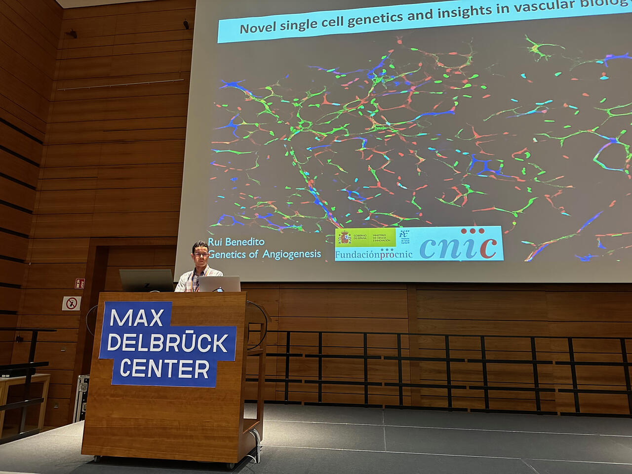
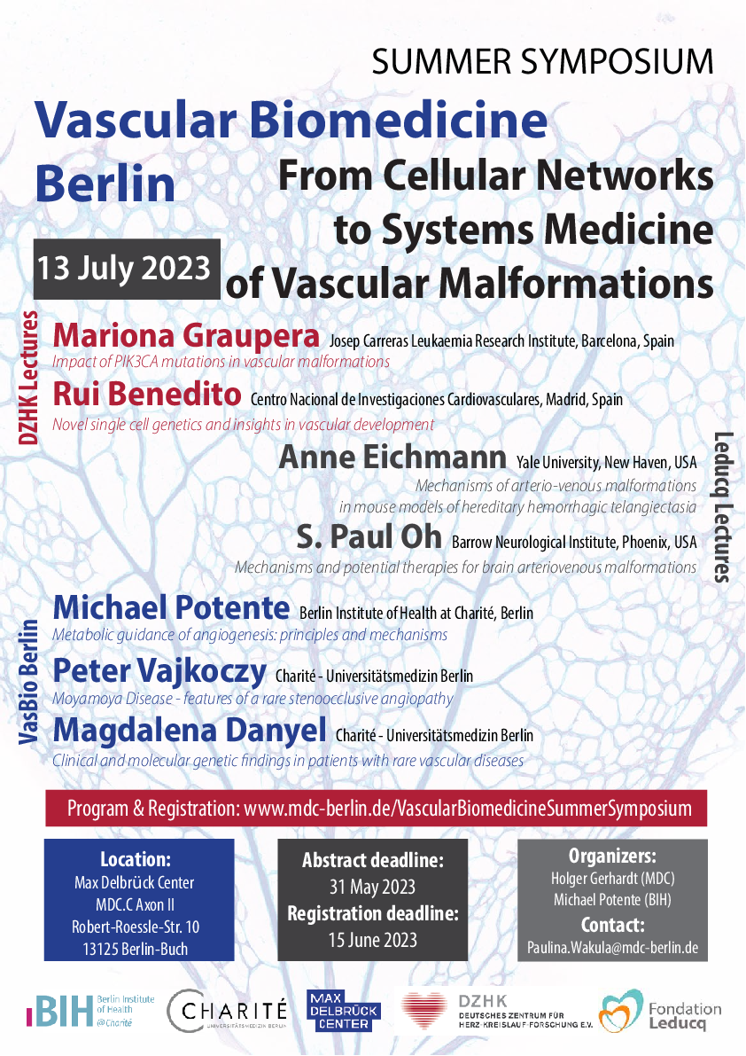
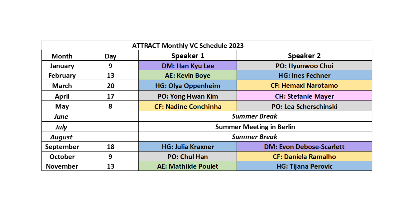


