A tricky kidney puzzle
To find out exactly what happens when in a particular cell, scientists look at its transcriptome – the totality of all genes that are expressed and transcribed into RNA at a specific point in time. Today, single-cell RNA sequencing allows the expression profiles of many thousands of cells to be analyzed simultaneously. But this method requires the cells to first be detached from their cell aggregate, which causes information on the cell’s original location in the tissue to be lost.
Nevertheless, gene expression enables this information to be reconstructed bioinformatically. “We wanted to know whether, in addition to providing a reconstruction of the cells’ spatial arrangement, algorithms could also be used to gain functional information from single-cell sequencing – for example, about the environmental conditions of kidney cells,” says Dr. Christian Hinze from Charité – Universitätsmedizin Berlin and the Molecular and Translational Kidney Research Lab at the Max Delbrück Center for Molecular Medicine in the Helmholtz Association (MDC). He is the first author of the study, which has now been published in the Journal of the American Society of Nephrology (JASN).
A heterogeneous organ
Over 15 different cell types are found in a mouse kidney, for example – and some are present in all three kidney regions.
Spatially speaking, the kidneys of mammals are very heterogeneous. The two bean-shaped organs consist of a renal cortex and an outer and inner renal medulla. The inner renal medulla is where highly concentrated urine forms after undergoing various filtering processes, and is then excreted via the ureter and bladder. “Over 15 different cell types are found in a mouse kidney, for example – and some are present in all three kidney regions,” says Hinze. Think it sounds like an impossible puzzle? Actually, it’s not. Because the microenvironment in which a cell lives is also reflected in its gene expression.
Back in 2019, the team led by Professor Nikolaus Rajewsky, Scientific Director of the MDC’s Berlin Institute for Medical Systems Biology (BIMSB) and one of the current study’s last authors, developed the program NovoSpaRc, which allows the spatial arrangement of cells within an organ to be reconstructed based on gene expression. In initial studies, the researchers found that cells that are arranged close to each other also resemble one another in terms of the activity of certain genes. “This is because they have to function in the same environment,” explains Hinze. “In the inner renal medulla, for example, conditions are much more severe than in the renal cortex. This is because the osmolality of the environment – i.e., the concentration of dissolved substances in the cell’s surroundings – increases drastically from the outside to the inside of the organ.”
An algorithm solves the 3D puzzle
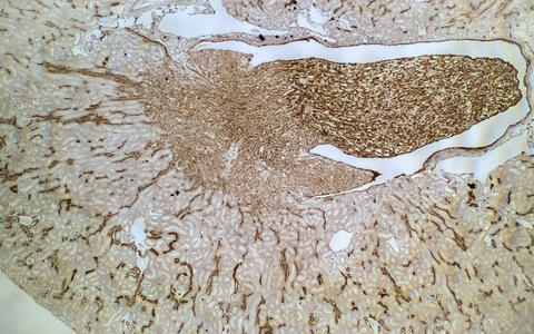
Section through a mouse kidney: The messenger RNA of a gene is stained brown. The more intense the staining, the stronger the gene expression. This illustrates the spatially heterogeneous expression of genes in the kidney.
For their current study, the researchers concentrated primarily on a specific cell type in the kidney known as principal cells. These cells play an important role in the reabsorption of blood salts and water and are distributed throughout the entire organ. Thanks to the particular gene expression of this cell type, they were able to extract these cells from the single-cell data of mouse kidneys. They then subjected the cells to the NovoSpaRc algorithm, which organized the expression data by comparing more than 800 selected genes for similarities. “NovoSpaRc works like someone doing a jigsaw puzzle,” explains bioinformatician Dr. Nikos Karaiskos from the Rajewsky Lab. “It tries to bring the different parts, which are the cells, together in such a way that the end result makes sense.” Karaiskos is also co-developer of NovoSpaRc and leading the DFG-funded “Unbiased Single-Cell Spatial Transcriptomics” project.
And the algorithm succeeded in completing the puzzle. “The gene expression analysis confirmed that osmolality in the tissue greatly increases along the axis from the renal cortex to the inner medulla,” says Hinze. “At higher salt concentrations, the cell switches on certain protective genes so that it can survive in this environment.” The researchers verified their results by carrying out comparative tests on tissue from genetically modified mice, in which the normal salt gradient had been disrupted.
An atlas of gene expression in the kidney
So, to what extent does spatial single-cell analysis actually advance kidney research? “We are now able to more accurately predict the spatial gene expression in the kidney, and thus also to make deductions about functions or malfunctions in certain regions of the kidney,” explains Professor Kai Schmidt-Ott, a nephrologist at the MDC and Charité and co-last author of the study. “We were already able to take a first step and create a high-resolution spatial atlas of the gene expression of healthy mouse kidneys, which we are now making available online to the scientific community.”
In one minute, the algorithm scans the data of a couple of thousand cell and for a full reconstruction of the small mouse kidney, it only needed a few seconds.
Previous data analyses are limited to healthy and genetically modified kidneys from animal models. Now, the researchers want to turn their attention to sick kidneys. “We believe that the new methods can also give us a better understanding of the regional molecular processes in kidney disease,” says Schmidt-Ott.
Reconstructed in seconds
Combining single-cell RNA sequencing with bioinformatic tissue reconstruction has a number of benefits. For one, several experiments are usually necessary to study different regions of an organ. “Now, we can simply cut up the entire kidney, sequence it, and determine all sorts of information from the transcriptomes – which saves a lot of time and money on research,” Hinze emphasizes. It can be used to investigate practically everything that is reflected in the gene expression of a cell – complete with spatial resolution. This includes the oxygen or nutrient supply of the tissue, or, as is the case here, the osmolality. What is particularly exciting, however, is that existing archived data sets can be used to answer new research questions – even if samples of the corresponding cells have long since ceased to exist.
And one last benefit: It takes practically no time for the expression data puzzle to be completed. “In one minute, the algorithm scans the data of a couple of thousand cells,” says Karaiskos. “And for a full reconstruction of the small mouse kidney, it only needed a few seconds.”
Text: Catarina Pietschmann
Further information
- Atlas of gene expression in healthy mouse kidneys
- Collaborative Research Center 1365 Renoprotection
- Press release: “3D maps of gene activity”
- Single-cell analysis at the MDC
Literature
Christian Hinze et al. (2020): „Kidney Single-cell Transcriptomes Predict Spatial Corticomedullary Gene Expression and Tissue Osmolality Gradients“. JASN, DOI: 10.1681/ASN.2020070930.
Downloads
Section through a mouse kidney: The messenger RNA of a gene is stained brown. The more intense the staining, the stronger the gene expression. This illustrates the spatially heterogeneous expression of genes in the kidney. Photo: Schmidt-Ott Lab, MDC.
Contacts
Christina Anders
Editor, Communications Department
Max Delbrück Center for Molecular Medicine in the Helmholtz Association (MDC)
+49-(0)30-9406-2118 christina.anders@mdc-berlin.de or presse@mdc-berlin.de
Dr. Christian Hinze
Molecular and Translational Kidney Research Lab
Max Delbrück Center for Molecular Medicine in the Helmholtz Association (MDC)
+49-(0)30-9406-2633 christian.hinze@mdc-berlin.de
- The Max Delbrück Center for Molecular Medicine (MDC)
-
The Max Delbrück Center for Molecular Medicine in the Helmholtz Association (MDC) is one of the world’s leading biomedical research institutions. Max Delbrück, a Berlin native, was a Nobel laureate and one of the founders of molecular biology. At the MDC’s locations in Berlin-Buch and Mitte, researchers from some 60 countries analyze the human system – investigating the biological foundations of life from its most elementary building blocks to systems-wide mechanisms. By understanding what regulates or disrupts the dynamic equilibrium in a cell, an organ, or the entire body, we can prevent diseases, diagnose them earlier, and stop their progression with tailored therapies. Patients should benefit as soon as possible from basic research discoveries. The MDC therefore supports spin-off creation and participates in collaborative networks. It works in close partnership with Charité – Universitätsmedizin Berlin in the jointly run Experimental and Clinical Research Center (ECRC), the Berlin Institute of Health (BIH) at Charité, and the German Center for Cardiovascular Research (DZHK). Founded in 1992, the MDC today employs 1,600 people and is funded 90 percent by the German federal government and 10 percent by the State of Berlin.


