Our group carries out interdisciplinary imaging projects using small animal and whole-body human MR scanners with high and ultrahigh field strengths to explore organs and tissues in novel ways. One of our aims is to support new approaches to diagnosis and therapy.
Magnetic resonance imaging (MRI) is a crucial diagnostic tool in clinics worldwide. The approach uses magnetic fields and radio frequency (RF) signals to depict tissues and organs. Research applications are rapidly expanding thanks to constantly increasing magnetic field strengths, customized RF antenna arrays, and faster, smarter image processing. Higher spatial resolution combined with the capacity to image new substances noninvasively is providing new insights into healthy and pathological processes under in vivo conditions.
Bringing MRI to nano-scale probes
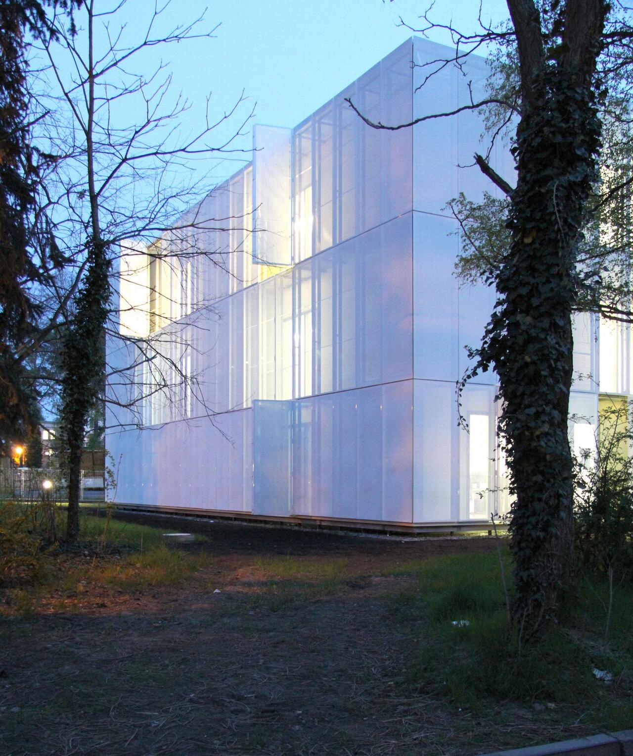
The Berlin Ultrahigh Field Facility (B.U.F.F.) at the Max Delbrück Center provides advanced MRI capabilities for interdisciplinary research projects using small animal and whole-body human MR scanners. We carry out human MRI on 7‑Tesla and 3‑Tesla instruments, and animal MRI at 9.4 Tesla. Significant gains in field strengths are enabling us to image new substances and nano-scale probes. We are also expanding imaging to new organ and model systems by custom-designing novel types of MR detectors (RF antennae).
The facility has reception areas and changing rooms for volunteers, and all the technical prerequisites for clinical studies, including emergency equipment and extra patient monitoring units. This has allowed us to take on an important role in major research initiatives, including the Helmholtz Imaging and Curing Environmental Metabolic Diseases (ICEMED) Alliance, the German National Cohort, and a number of other national and international projects devoted to various health conditions.
Mapping the heart and kidney in new ways
B.U.F.F. is collaborating with other research groups to develop new methods for mapping anatomy, morphology, microstructure, function, physiology, and metabolism in animal and human subjects. By achieving new levels of spatiotemporal resolution, and imaging new substances such as sodium, we are able to conduct groundbreaking studies on organs such as the heart and kidney. We have begun a new thermal phenotyping project, supported by the European Research Council (ERC), to characterize the temperature profiles of various tissues, both healthy and diseased. One aim of the project is to use MRI to manipulate the temperature of tissues and utilize this parameter as a potential diagnostic and therapeutic tool.
Principle investigators of B.U.F.F. form a key/integral part of major initiatives including the Helmholtz Alliance for Imaging and Curing Environmental Diseases (ICEMED), the population MRI program of the German National Cohort (GNC), the DFG research group FOR1368 on Hemodynamics of Acute Kidney Injury, the imaging program of the German Center for Cardiovascular Research (DZHK), the EU funded FP7 project INSERT on hybrid SPECT/MR imaging, the BMBF initiative on sodium MRI (NAMRIS) and the German Ultrahigh Field Imaging (GUFI) network. B.U.F.F. is an integral part of the imaging core facility of the Berlin Institute of Health (BIH).
Safety instructions
Research Opportunities
The Berlin Ultrahigh-Field MR Facility at the Max-Delbrück-Center for Molecular Medicine (MDC), Berlin, Germany is seeking enterprising young scientists interested in the basic development and clinical application of ultrahigh-field magnetic resonance imaging (UHFMRI) as:
Diploma‑, Master- and Bachelor Thesis Projects or Internships in Ultrahigh-Field MR Imaging (pdf)
PhD Thesis
Following Immune Cell Therapies In Vivo by Magnetic Resonance Methods (pdf)
Investigation of in vivo drug distribution by Magnetic Resonance Methods (pdf)
Ultrasensitive Fluorine Magnetic Resonance Imaging (pdf)