📺 Like a deep-sea diver
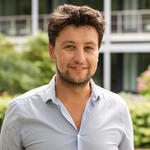
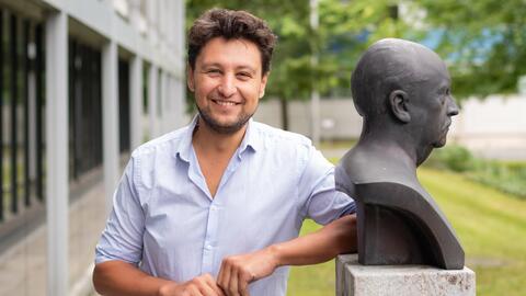
Dr Misha Kudryashev describes his entry into the world of biophysics as a ”dive into the unknown”. 16 years ago, he left his native Russia to start this academic research in Germany. In the years since he has settled in – not only in biophysics but also in his new home. Since 1 August, the 38-year-old has been establishing a research group at the Max Delbrück Center for Molecular Medicine in the Helmholtz Association (MDC) dedicated to “In situ Structural Biology”.
His main research interest is the structural biology of membrane protein complexes. These are proteins embedded in the lipid layers of cell membranes. They play an important role in the life of cells, one if the mechanism is the activation of ion channels, which are the main modulators of signalling in nerve and muscle cells. “I want to understand how these membrane proteins are structured and regulated,” says Misha Kudryashev. “Their control mechanisms are very interesting from the perspective of drug development. Substances with the ability to switch specific proteins on or off can cure diseases or improve the quality of life.”
Cryo-EM provides exceptionally high-resolution

The cryo-EM team (from left to right): Dr. Christoph Diebolder, Dr. Thiemo Sprink and Metaxia Stavroulaki in front of the four-meter-high cryo-electron microscope.
To dive into the hidden world of proteins, their structure and their mechanisms of action, Kudryashev uses modern imaging techniques such as cryo-electron microscopy (cryo-EM) and cryo-electron tomography (cryo-ET). These techniques make it possible to examine cells or their individual components in their natural state and in their cellular environment – “in situ”. For cryo-EM purified proteins or entire cells are quickly frozen to very low temperatures. An electron beam penetrates the sample, which is encased in a thin layer of vitreous, non-crystalline ice. This produces transmission images that are recorded. Many two-dimensional images of a sample are converted by software into a precise three-dimensional representation known as a tomogram. Tomograms of cells provide information about the interactions of molecules inside cells and about structure of proteins “in situ”, i.e. in their native environment. Kudryashev’s team develops algorithms for this technically challenging process to achieve the highest possible resolution.
For this work, he benefits from the outstanding infrastructure at the research campus in Buch. In March of this year, the Cryo-EM Core Facility at Charité – Universitätsmedizin Berlin started work in partnership with the MDC and the Leibniz-Forschungsinstitut für Molekulare Pharmakologie (FMP). Misha Kudryashev is full of enthusiasm for the excellent equipment and the cryo-EM team: “The team really knows their stuff. I thoroughly enjoy working with them.”
“Something to do with computers“
Misha Kudryashev ended up in biophysics by accident – like a diver swimming in the deep without knowing what awaited him, but then discovering something that determined the direction his life would take. He originally intended to study mathematics but didn’t pass the entrance exam. Because he wanted to do “something with computers” and physics involves a lot of data, he decided to study physics at the University of Krasnoyarsk in Russian Siberia. At the same time, he worked as an early career researcher at the Institute of Computational Modelling, where he analysed the statistics of protein sequences for his Diploma thesis. It was 2001 and the human genome had just been decoded. “I was fascinated by the big data produced by protein sequencing, and the potential to predict structures of proteins from their sequences” he recalls. In 2005, he graduated with distinction.

Dr. Misha Kudryashev, In situ Structural Biology Lab
“After I finished my undergraduate degree, going abroad was really the only option,” the researcher explains. “I knew that I wanted to go into research, but at the time I couldn’t have done that in Russia without having an additional job”. He applied for a PhD position at the European Molecular Biology Laboratory (EMBL) in Heidelberg, however, again, failed an entry exam, this time in biology. Nonetheless, he received an offer to start working in another Heidelberg group at more or less the same time. The group led by Professor Friedrich Frischknecht at the University of Heidelberg, investigates how malaria parasites manage to pass unharmed from a mosquito through the insect’s proboscis into human skin and then actively travel through the skin into a blood vessel. In the search for a drug that would stop the motility of the parasites, Misha Kudryashev’s job focused on image analysis for the drug screen. Using fluorescence microscopy, the researchers quantified how the chemical compounds affected the motility of malaria parasites. They succeeded faster than expected – and, as if by chance, they were able to image the architecture of the unicellular parasites with the help of cryo-electron microscopy. The team had an access to one of the first specialized microscopes installed at the Max Planck Institute of Biophysics in Munich – at least when it wasn’t used by other researchers, recalls Kudryashev. “I spent many nights in Munich recording data instead drinking beer.”
The technology, combined with computer-assisted data processing, fascinated him. In 2009, after four years in Heidelberg, Misha Kudryashev joined the Biozentrum at the University of Basel. In the lab headed up by Professor Henning Stahlberg, he studied proteins in the membranes of bacteria. He was primarily interested in how bacteria excrete enzymes and toxins into their environment or the host cells. He investigated the secretion system used by the pathogens that cause plague (Yersinia pestis) and dysentery (Shigella) and produced the first structures of this protein complex from cryo electron tomograms, enhancing their resolution. “It’s a very effective method,” he says, “we still use it now”.
“There’s a lot involved in structural biology”
I believe that the structures of membrane proteins as we understand them at the moment are not all quite correct. Or rather, I hope they aren’t – then my research will be more meaningful.
Misha Kudryashev took charge of his own group for the first time in 2015 at the Max Planck Institute of Biophysics and the Buchmann Institute for Molecular Life Sciences at Goethe University in Frankfurt am Main. Funded by the Sofja Kovalevskaja Award from the Alexander von Humboldt Foundation and by the German Research Foundation (DFG), he and his team examined the structure and activation of ion channels. His research in Berlin follows on seamlessly from his previous work. “We want to unravel the structure of membrane proteins in native context, improve the methods available to do this, and make these methods available to the research community.” He plans to establish long-term research links and create a supportive environment for fellow researchers. “There’s a lot involved in structural biology,” says the physicist. “The hardware is expensive and the software is complicated. The more people that get involved, the easier it is for other researchers to become part of the network.”
In the long term, he wants to achieve a deeper understanding of membrane protein complexes – how they are structured, how they function and how they can be influenced. ”I believe that the structures of membrane proteins as we understand them at the moment are not all quite correct. Or rather, I hope they aren’t – then my research will be more meaningful.”
Text: Jana Ehrhardt-Joswig