New clues on how the heart makes arteries
A team led by Dr. Elena Cano in the Integrative Vascular Biology Lab of Professor Holger Gerhardt at the Max Delbrück Center in Berlin has elucidated the mechanism via which new arteries form in the heart. The finding, published in “Circulation Research,” fills in an important gap in our understanding of how coronary arteries develop and could help improve treatments that aim to heal damage to heart muscle caused by a heart attack or stroke – leading causes of death and disability worldwide. The work was supported by the DZHK (Deutsches Zentrum für Herz-Kreislauf-Forschung).
A heart attack or stroke can cause tissue within heart muscle to die. As a result, the blood supply to the heart diminishes, leading to permanent disability. Although some tissue does spontaneously heal by growing new blood vessels, the extent of is often inadequate to completely restore blood supply. Current therapies focus on managing symptoms and slowing heart disease.
Researchers have attempted various methods to coax new blood vessels to grow in damaged heart tissue. However, they have not been able to establish a stable, mature vascular network that improves the heart’s function, says Cano. One major drawback has been the lack of deeper understanding of the intricate molecular, cellular, and structural cues that vascular cells use to build a hierarchical blood vessel network.
Applying single-cell sequencing to the problem
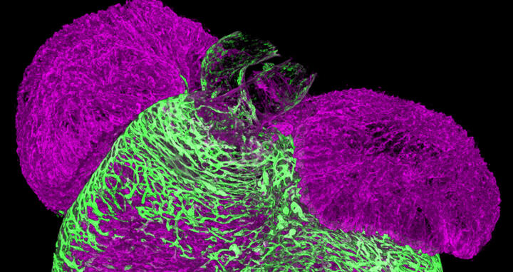
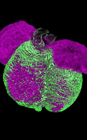
A 3D image of an embryonic mouse heart. Endothelial cells vascularize the heart muscle walls (shown in green). This process establishes the coronary vasculature that nourishes the heart muscle.
While studying how tissues vascularize, Cano discovered a type of pre-arterial cell in her samples that appeared to play a major role in growing new arteries. These pre-arterial cells had already been reported by other researchers. But Cano wanted to study them with new technology.
Using single-cell sequencing to analyze the transcriptome of cardiac cells taken from mice at different stages of development, the team of researchers showed that these cardiac pre-arterial cells develop from “tip cells” – specialized cells that sense environmental cues to drive growing vessels into specific directions. The team also use 3D spatiotemporal mapping to confirm their results. Moreover, they showed that these pre-arterial cells were already “marked” to develop into arterial cells, which belies current thinking about how arteries develop.
It had been thought that new arteries form their unique characteristics, such as length and diameter, based purely on mechanical cues, such as the velocity of the fluid flowing through them. However, “this study shows that pre-arterial cells already have characteristics of arteries before any fluid flows through them,” Cano says.
The researchers also reanalyzed previously published single-cell data collected by researchers in the U.K. from human embryonic heart tissue. They then compared it to their own data collected from mouse heart tissue damaged by heart attack. They found that new arteries formed in the human embryonic tissue via the same mechanism as after damage from a heart attack in mice. “We show that not only is this mechanism conserved between mice and humans during development, but it also persists throughout life and becomes activated after a heart attack,” says Cano.
Implications for treatment
Understanding how coronary arteries naturally form and regenerate opens the possibility of developing treatments that stimulate these regenerative pathways, potentially reversing heart muscle damage, says Cano.
“Now we know that it is not only flow that signals vascular endothelial cells to become arteries, but that tip cells transition into pre-arterial cells to eventually become arteries,” says Gerhardt. “Surprisingly, we find that not all tip cells have the capacity to form arteries, raising the prospect of selectively increasing this pool of cells for therapeutic purposes.”
“This is a step forward,” Cano adds. “It is a new mechanism that we can potentially tweak during regeneration to see whether we can form new arteries for optimal restoration of blood supply.”
Text: Gunjan Sinha
Further information
Literature
Elena Cano, Jennifer Schwarzkopf, Masatoshi Kanda, et. al. (2024): “Intramyocardial Sprouting Tip Cells Specify Coronary Arterialization.” Circulation Research, DOI:10.1161/CIRCRESAHA.124.324868
Photo for download
Photo credit: Max Delbrück Center
Contacts
Prof. Holger Gerhardt
Group Leader
Integrative Vascular Biology Lab
Max Delbrück Center
Holger.Gerhardt@mdc-berlin.de
Gunjan Sinha
Editor, Communications
Max Delbrück Center
+49 30 9406-2118
Gunjan.Sinha@mdc-berlin.de or presse@mdc-berlin.de
- Max Delbrück Center
-
The Max Delbrück Center for Molecular Medicine in the Helmholtz Association (Max Delbrück Center) is one of the world’s leading biomedical research institutions. Max Delbrück, a Berlin native, was a Nobel laureate and one of the founders of molecular biology. At the locations in Berlin-Buch and Mitte, researchers from some 70 countries study human biology – investigating the foundations of life from its most elementary building blocks to systems-wide mechanisms. By understanding what regulates or disrupts the dynamic equilibrium of a cell, an organ, or the entire body, we can prevent diseases, diagnose them earlier, and stop their progression with tailored therapies. Patients should be able to benefit as soon as possible from basic research discoveries. This is why the Max Delbrück Center supports spin-off creation and participates in collaborative networks. It works in close partnership with Charité – Universitätsmedizin Berlin in the jointly-run Experimental and Clinical Research Center (ECRC), the Berlin Institute of Health (BIH) at Charité, and the German Center for Cardiovascular Research (DZHK). Founded in 1992, the Max Delbrück Center today employs 1,800 people and is 90 percent funded by the German federal government and 10 percent by the State of Berlin.


