
The cellular engineer
For Professor Oliver Daumke, a cell is like an engine room – molecular machines made up of protein building blocks are continuously at work. These protein machines transport nutrients into the cell or help neutralize unwanted invaders among other tasks. It is a precisely choreographed dance of countless miniature processes.
For most of his scientific career, Daumke has been peering into this engine room of life to understand the components of these molecular machines. Using X‑ray crystallography and cryo-electron microscopy, he deciphers the identities of the amino acids that make up chains of proteins of interest, and then investigates how these chains fold into 3D machines that are smaller than the wavelength of visible light.
“Like the blade of a pocketknife”
Opening his laptop, Daumke plays a short, animated film showing the choreography of a 3D model of GBP1 – a critical protein for the innate immune response in humans. GBP1 targets bacterial invaders like salmonella, latches onto them, encapsulates them, and disrupts their outer membranes so they can no longer replicate. “We discovered how the GBP1 machine positions itself on bacterial membranes to destroy them,” Daumke says. “Its protein structure changes dramatically when bound to bacteria and begins to form a uniform shell around the pathogen.”

The molecular choreography of GBP1 unfolds on the monitor in front of us, as it would take place in human cells following an infection. Two protein building blocks bind to two energy carrier molecules, called GTP, and connect head-to-head. Other proteins use the energy of GTP to open a molecular lever. “Like the blade of a pocket knife,” notes Daumke. They then combine with thousands of other unfolded GBP1 proteins to form what looks like spokes of a wheel, which in turn stack up with many other wheels to form tubes. These tubes attach themselves to the membrane of the bacterium – and make it permeable. Other, as yet unknown, immune defense molecules can then penetrate the bacterium and render it harmless.
Daumke’s eyes light up, as if seeing the animation for the first time. “That unfolding motion is absolutely spectacular,” he says. Understanding the molecular mechanism behind how GBP1 works fascinates him. While this is basic research, it has clinical implications – it could lead to drugs that boost the immune response to bacterial infections.
Small changes, major diseases – and paths toward cures
At the Max Delbrück Center, Daumke’s team is often pulled into projects that have found a faulty protein that is linked to disease. “Mutations in our genes can impair their function,” he says. “In the worst case, changing a single amino acid can cause a devastating illness.”

For example, when mutations make the dynamin protein overactive, it can lead to centronuclear myopathy – a fatal muscle disease. Vital for normal cell function, overactive dynamin disrupts muscle structure. Structure-function analyses help pinpoint which amino acids are responsible for such malfunctions — and how they might be corrected.
Daumke has spent 12 years studying the dynamin family of proteins, which play key roles in endocytosis – a process cells use to ferry substances inside. Partner proteins guide dynamin to the site on the cell membrane where, for example, iron molecules are to be taken up via a process called membrane invagination. Around 40 dynamin units then form a ring- like structure around neck of the invagination site. “Dynamin works like a ratchet, tightening the neck further and further until the invagination can be separated together with the iron molecules and then taken into the cell,” says Daumke. He first determined the structure back in 2011 and then created a comprehensive computer model of the process using his findings in 2021.
Such structural insights can have direct medical applications. Daumke’s team, for example, modified the structure of an antibody in such a way that it could be injected. The antibody is currently being tested in patients with multiple myeloma – a type of incurable bone marrow cancer. “The antibody is still in clinical trials, but in one patient, the cancer has completely disappeared,” Daumke says. The antibody could serve as the basis of a new therapy.
A life in service of protein machines
Asked what defines him as a scientist, Daumke points to persistence, an obsession with detail, and a willingness to challenge his own knowledge. Those qualities may explain his long-standing fascination with the highly complex science of protein structure and function.

As a student at the University of Cologne, he studied a transporter protein that moves peptides across membranes. For his doctorate at the Max Planck Institute of Molecular Physiology in Dortmund, he determined the 3D structure of Rap1GAP, and discovered a completely new mechanism through which the protein switches off a molecular signal in cells. During his postdoc at the Laboratory of Molecular Biology in Cambridge – a cradle of structural biology – he unraveled the workings of EHD2, a protein that forms a ring on cellular membranes to help to stabilize pores within the cell.
Daumke joined the Max Delbrück Center in 2007 as head of a junior research group. Since 2013, he has been Senior Group Leader of the Structural Biology of Membrane-Associated Processes lab. He also holds a professorship at Freie Universität Berlin. Today, his team includes 15 researchers.
Seeing the whole cell
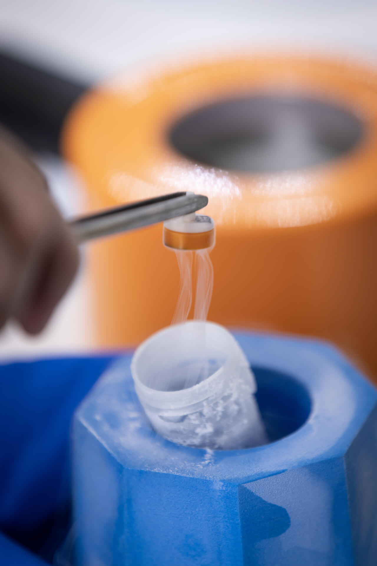
Cryo-electron microscopy, which won the 2017 Nobel Prize in Chemistry, has greatly accelerated structural biology research. The technology allows scientists to visualize highly complex proteins and their spatial arrangement. Samples are flash-frozen in liquid nitrogen and imaged from multiple angles with electron beams, producing detailed 3D reconstructions. Daumke’s lab has been using this technology since 2017.
For a long time, structural biologists had been taking the approach of reducing complexity by looking at proteins in isolation using X‑ray crystallography. Cryo-EM opened the door to studying proteins in their native cellular environment. Daumke’s team also combines the method with light microscopy to tag proteins and locate them inside cells.
“Many proteins are dependent on other cellular partners or membranes to function,” he says. “To understand how these protein machines really work, you have to look at the whole picture.” This approach is known as integrative structural biology. The ability to view an entire cell, also enables scientists to better understand the effects of genetic mutations and address them therapeutically.
AI-powered predictions
The second big change in Daumke’s field is artificial intelligence. Google’s AlphaFold platform has calculated the structure of 200 million proteins in three-dimensional space and made them freely accessible to the scientific community. “This means that it is often no longer necessary to determine structures ourselves,” says Daumke. What can take several years in the laboratory, AlphaFold does in just a few minutes. However, many questions can still only be answered experimentally: how proteins are modified after they are synthesized, for example, how they interact, or how they move.
But this does not mean that structural biologists are becoming obsolete – quite the opposite. Combining integrative methods with AI is making it possible to answer entirely new questions. The ultimate goal? A full simulation of a cell with all its moving, interlocking parts. “How an entire cell works, in which thousands of protein machines work simultaneously, and how it is all coordinated, is a big question. Many disciplines must come together to solve this,” says Daumke. He wants to make a contribution by using the latest technologies to look even deeper into the engine room of the cell and trace the miniature processes of life – cog by cog.
Text: Mirco Lomoth


