Two first-place awards in scientific image contest
Every neuron in the brain is connected to thousands of others via synapses, and these cells communicate using neurotransmitters. A team led by Dr. Jana Kroll from Professor Oliver Daumke’s “Structural Biology of Membrane-Associated Processes” lab has captured this process in an image titled Excitation. This image has now been awarded the Jury Award in the “Best Scientific Image Contest,” organized annually by the Helmholtz Imaging.
Out of 104 entries, an independent jury of international experts in science and art selected the image, created using cryo-confocal microscopy, for the award. Three other images from researchers at the Max Delbrück Center are also among the top 20 and will be featured in a traveling exhibition across various Helmholtz centers. The images will be displayed as 50 x 70 centimeter prints.
The other highlighted images were created by Athanasios Balomenos, Dr. Wenhan Luo, and Professor Gary Lewin from the “Molecular Physiology of Somatosensory Perception” research group; Ariana Rauch from the “Immuno – Microbial Dynamics in Cardiorenal Disease” research group led by Dr. Nicola Wilck; and Dr. Andreas Marg from the “Myology” research group led by Professor Simone Spuler at the Experimental and Clinical Research Center (ECRC), a joint institution of Charité – Universitätsmedizin Berlin and the Max Delbrück Center.
The dance of the starfish embryos
In addition to the €1,000 Jury Award, Helmholtz Imaging also presents two other awards every year: the Public Choice Award, where the Helmholtz community votes for their favorite image, and a Participants’ Choice Award, chosen by attendees at the Helmholtz Imaging Conference. The conference, where competition winners were officially announced, took place in Potsdam from June 25 to 27.
This year, Dr. Rafael Deliz Aguirre received the Public Choice Award and a cash prize of €600. His image, Starfish Puts Motion Back in the Ocean, was assembled during his time in the Spatial Proteomics lab led by Dr. Fabian Coscia. It shows starfish embryos, whose microscopic tentacles move to generate currents that help propel them, and make the dynamics of their movements visible.
First place in the Participants’ Choice Award is shared by two teams. Aldino Rizaldy, Dr Sam Thiele and Dr Sharad Kumar Gupta from the Helmholtz-Zentrum Dresden-Rossendorf (HZDR) have created a digital twin of a forest to estimate its biomass – an increasingly important task in climate research. Their image is called Digital Forest / Digital Twin. Jenny Hein and Dr Thomas van de Kamp from the Karlsruhe Institute of Technology (KIT) impressed the conference participants with their photo A Bark Beetle’s Stellar Gut. The 3D synchrotron scan reveals the star-shaped foregut of a bark beetle. The image Filming the Smelling Brain by Balomenos, Luo and Lewin from the Max Delbrück Center came third in this category. Luo received the prize on behalf of his team at the Potsdam conference.
First Place, Jury Award
Excitation – Dr. Jana Kroll, Daumke Lab
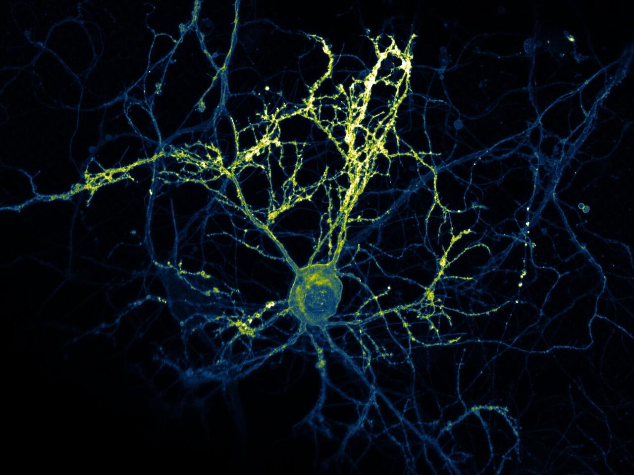
This image captures a mouse hippocampal neuron surrounded by numerous extensions that connect to other neurons via synapses. The cultured cell is equipped with a yellow fluorescent sensor for the neurotransmitter glutamate, which is normally used at room temperature. In this case, the image was captured at ‑170 °C using CFcryo-EM (Core Facility for Cryo – Electron Microscopy).
“We first stimulated individual neurons and then froze them just milliseconds later, capturing the precise moment the cell released its messenger substance,” explains Kroll. What’s special about this image is that the nerve cell exhibits dendrites with higher and lower fluorescence intensities. “This proves that both the timing and strength of signal transduction can vary significantly within a single neuron.”
First Place, Public Choice Award
Starfish Puts Motion Back in the Ocean – Dr. Rafael Deliz Aguirre, Coscia Lab
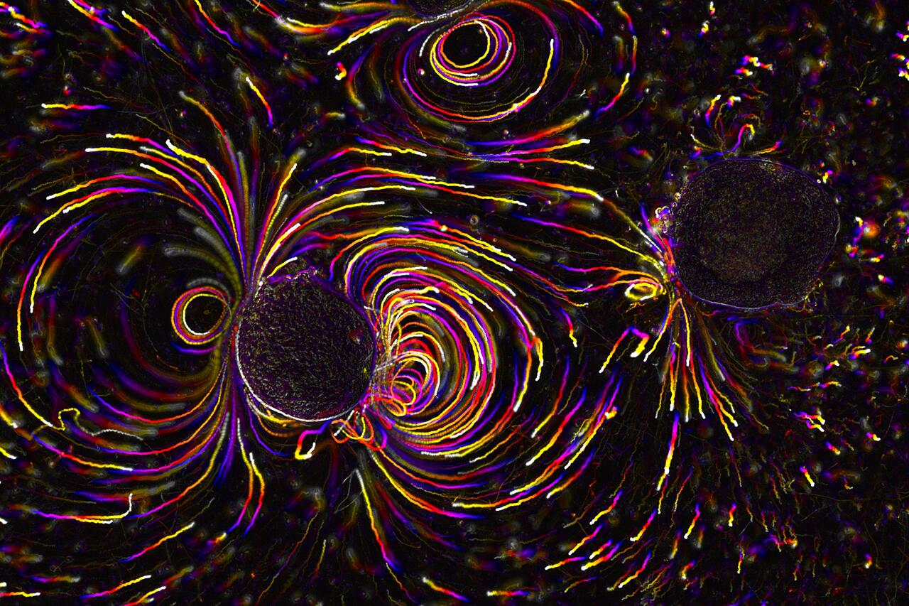
This image shows starfish embryos that are only a few days old, using their tiny tentacles to create currents that visually and mathematically reflect the Earth’s magnetic field. The living embryo on the left of the image – unlike the immobile, dead embryo on the right – is actively shaping its fluid environment. The image was captured using bright-field microscopy and fluorescent particles.
“The technique involved capturing several images over time, color-coding them, and merging them into a single picture,” explains Deliz Aguirre. The result is a dynamic snapshot that captures how starfish embryos move through the ocean. “The swirling, vortex-like patterns can be described with equations that could also apply to black holes,” says Deliz Aguirre. “The image reveals a deep link between microscopic and celestial physics.”
Among the top 20 jury selections
Filming the Smelling Brain – Athanasios Balomenos, Dr. Wenhan Luo, and Prof. Gary Lewin, Lewin Lab
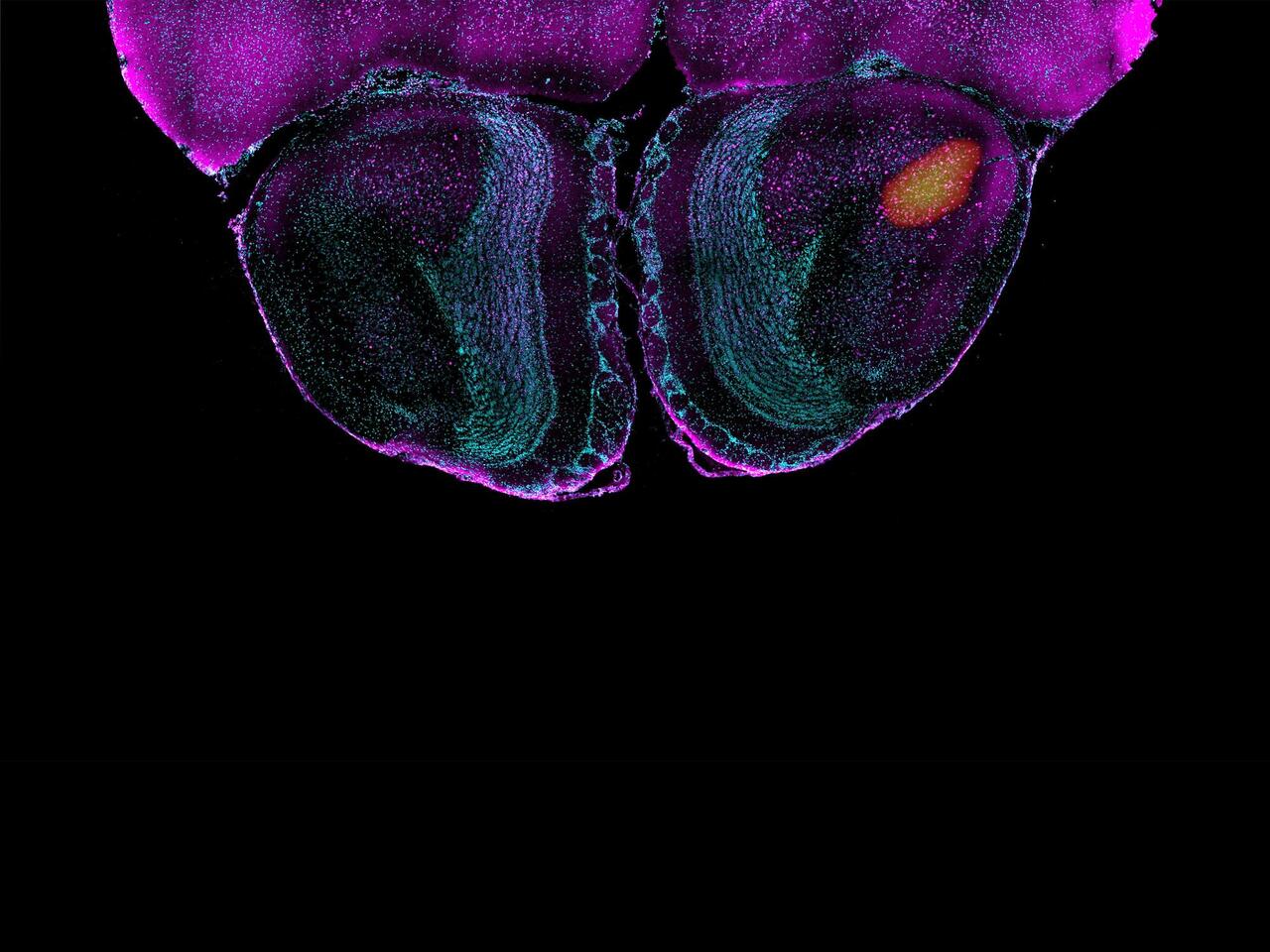
This image shows how specific stimuli activate neurons in the olfactory centers of mouse brains in real time. Created through a combination of functional ultrasound imaging (fUSI) and high-resolution microscopy, the image focuses on the anterior olfactory nucleus (AON), a critical part of the olfactory cortex that plays an important role in processing odors but has been little studied until now. “Our results suggest that the AON responds selectively to certain stimuli, providing new insights into its role in the sense of smell,” says Balomenos.
The Immune Landscape of the Gut – Ariana Rauch, Wilck Lab

This image captures a mouse colon, with T‑cells (yellow) and other immune cells (red) located near or within the epithelium (gray) that separates the intestine from its internal microbiome. The nuclei of the cells are shown in blue. “We investigated the position and distribution of these immune cells and their interaction with the microbiome,” explains Rauch. “Our goal is to identify subgroups influenced by the gut flora, shedding light on how changes in the microbiome affect immune responses in the intestine during disease.” The researcher captured the image of the stained cryosection using a fluorescence microscope.
Healthy Muscle After Gene Editing – Dr. Andreas Marg, Spuler Lab
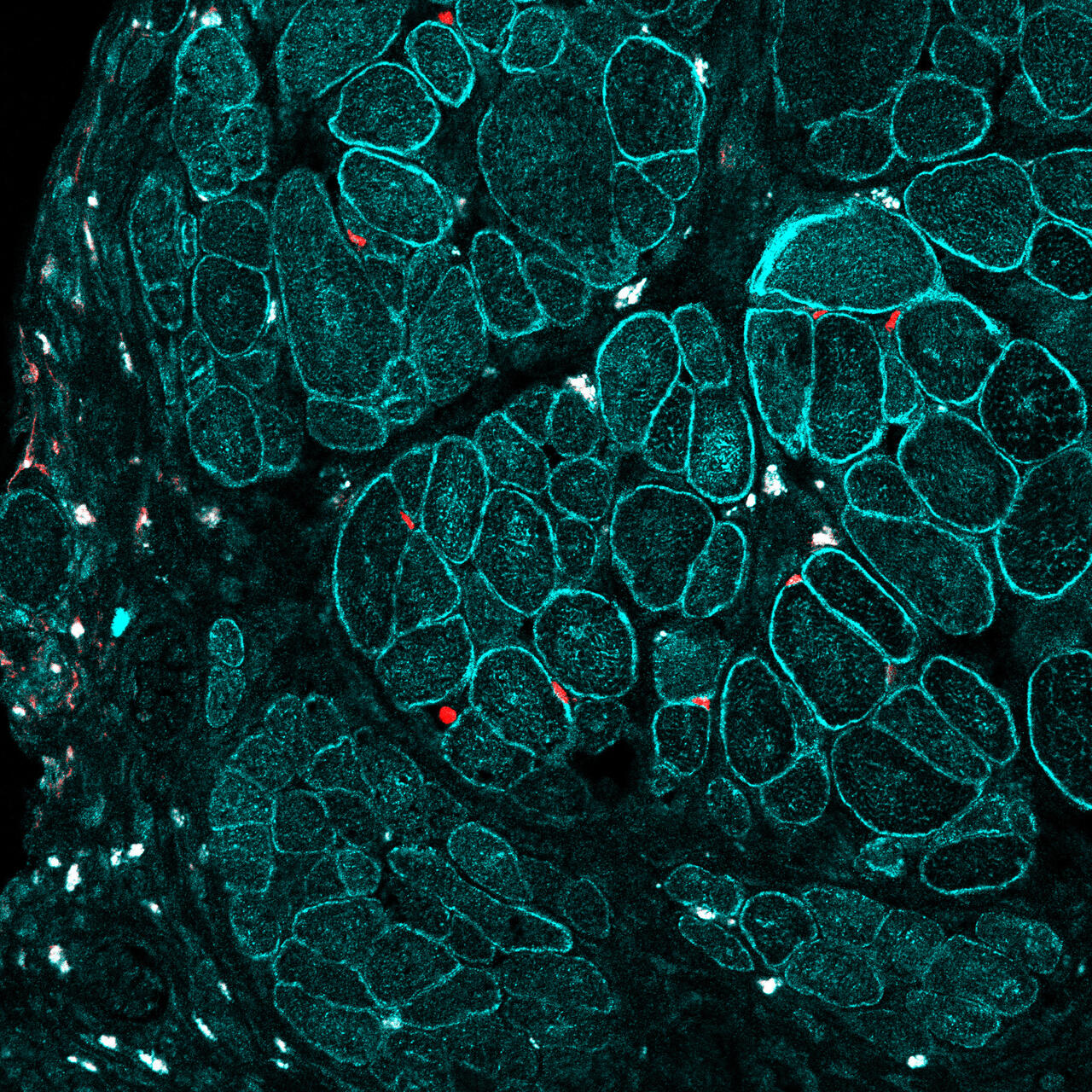
This image shows regenerated muscle tissue from a genetically modified mouse. Three weeks earlier, the researchers injected muscle stem cells in which a human disease-causing mutation had been corrected using the CRISPR/Cas9 gene-editing tool. The newly formed muscle fibers express the protein dysferlin (turquoise), while satellite cells (red) express the transcription factor Pax7, critical for muscle development and growth. “The results demonstrate that genetically modified cells can restore muscle integrity in diseased tissue,” says Marg. “This is a promising step toward treating dysferlin deficiency, an as-yet incurable form of muscular dystrophy.”
Text: Anke Brodmerkel