
What a waste! The molecular causes of muscle wasting in heart failure
New molecular details of why heart failure patients suffer from muscle wasting have been revealed by researchers at the Experimental and Clinical Research Center in Berlin and their collaborators at the University of Texas Southwestern Medical Center in Dallas.
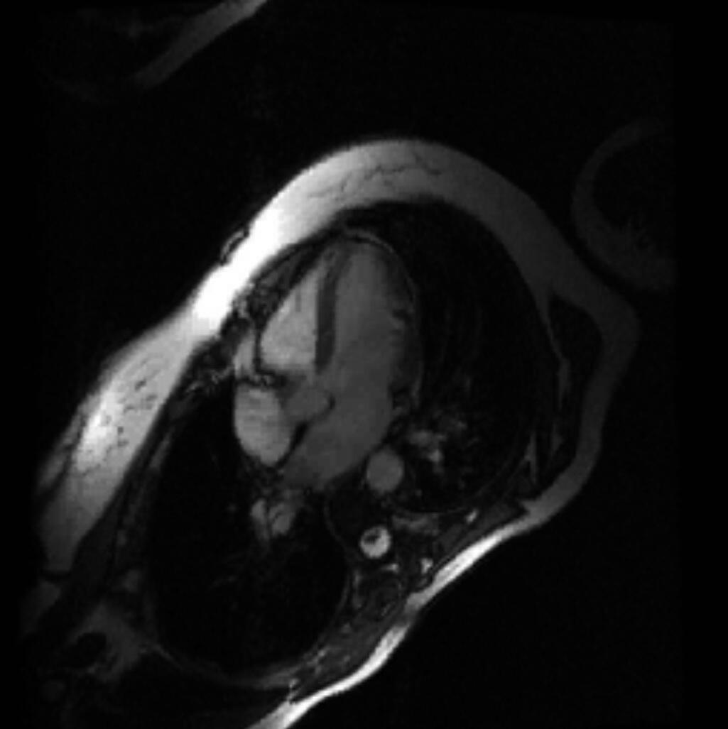
“Muscle loss is common in patients with end stage congestive heart failure and it’s associated with a poor prognosis,” explains clinician researcher Dr. Jens Fielitz who led the study. “We can provide treatment that improves the prognosis for heart failure patients, but in advanced stages of the disease they lose a lot of weight which worsens their condition”.
The weight loss is largely caused by muscle wasting, which happens when patients have high levels of the protein Angiotensin II in their blood. When Angiotensin II is high, the enzyme Muscle RING Finger 1 (MuRF1) is activated and tags muscle proteins for breakdown by the ubiquitin proteasome system – a cellular shredding machine that regulates protein degradation and turnover.

To treat heart failure, doctors prescribe drugs to inhibit the renin-angiotensin-aldosterone system such as Angiotensin Converting Enzyme (ACE) inhibitors and angiotensin II receptor blockers. As a positive side effect, these drugs also prevent muscle proteins from being broken down. “These drugs are part of the standard treatment for heart failure,” says Fielitz. “Both drugs reduce muscle loss, but only for 5 – 10 years of treatment when they lose their effectiveness.”
To develop new treatments against muscle wasting, it’s important to understand the molecular links between Angiotensin II and MuRF1. Fielitz and his team set out to study the pathway in detail.
In muscle atrophy, MuRF1 levels are 40 – 60 fold higher than normal, leading to the breakdown of muscle proteins, muscle loss and weakness. The researchers looked for transcription factors that could be responsible for the high MuRF1 levels behind muscle loss in heart failure patients. “Some transcription factors that control MuRF1 expression were known, but their activity was too weak to account for the large increase in MuRF1 levels we see in muscle atrophy,” Fielitz explains.
To seek regulators of MuRF1 the scientists screened a library of genes expressed in muscle cells. They used the fluorescent reporter gene luciferase behind the MuRF1 promoter. This gave a fluorescent indication of which genes encoded transcription factors activating MuRF1.
One gene stood out in the screen – transcription factor EB (TFEB). It was a strong activator of the MuRF1 promoter, capable of increasing MuRF1 levels by 70 – 100 fold. This was consistent with the search for a transcription factor that could cause large increases in MuRF1 in heart failure induced muscle wasting.
Further experiments confirmed TFEB could regulate MuRF1 expression in muscle cells. The researchers found that treating cells with Angiotensin II increased TFEB binding to the MuRF1 promoter. This demonstrated that the Angiotensin II pathway known to cause muscle wasting in heart failure regulates TFEB activity.
Next Fielitz and his team looked for links between Angiotensin II and TFEB. They found two proteins are important in this pathway, the histone deacetylase HDAC5 and the protein kinase PKD1.
HDAC5 acts as the brakes on muscle protein breakdown. It does this by binding TFEB so that it can’t activate MuRF1 expression. For HDAC5 to work as a brake it needs to be in the nucleus where it can bind TFEB. PKD1 releases the brakes by phosphorylating HDAC5, which sends it out of the nucleus and leaves TFEB free to upregulate MuRF1.
The researchers suspected that Angiotensin II triggers muscle wasting by activating PKD1, so they tested this theory in a mouse model. They used mice with a conditional knockout for PKD1 in their muscle cells to see how their muscles reacted to Angiotensin II.
Two groups of mice took part in the Angiotensin II trials, mice lacking PKD1 in muscle and control mice. The control mice experienced muscle loss in response to the Angiotensin II treatment and tests showed that they had increased MuRF1 levels and TFEB bound to the MuRF1 promoter. In contrast, the mice that lacked PKD1 in muscle cells were protected from the effects of Angiotensin II. Without PKD1 to release the brakes on MuRF1 expression, their muscles were not affected by the treatment.
The studies by Fielitz and his team have revealed the molecular details of the pathway between Angiotensin II and MuRF1-mediated muscle loss. This knowledge provides potential new targets for drug research aimed at preventing muscle wasting in heart failure.
“We hope that knowledge of this pathway and the potential drug targets within it will lead to new treatments for muscle wasting in patients with advanced congestive heart failure,” said Fielitz.
- Arwen Cross
Highlight Reference:
Du Bois P., Pablo Tortola C., Lodka D., Kny M., Schmidt F., Song K., Schmidt S., Bassel-Duby R., Olson E.N., Fielitz J. (2015). Angiotensin II Induces Skeletal Muscle Atrophy by Activating TFEB-Mediated MuRF1 Expression. Circulation Research 117: 424 – 436 (August 2015). DOI: 10.1161/CIRCRESAHA.114.305393
More about the Fielitz research group at the MDC/ECRC


