openDVP makes spatial proteomics broadly accessible
Today’s commercial satellites can image landscapes with a level of detail unthinkable 20 years ago: they capture individual trees, cracks in infrastructure and even details of road signs. In proteomic research, Deep Visual Proteomics (DVP) enables scientists to do something similar at the cellular and molecular scale — they can zoom in on selected cells and cellular communities and measure thousands of proteins while preserving information about where those cells are located in a tissue.
Now researchers in the Spatial Proteomics lab headed by Dr. Fabian Coscia at the Max Delbrück Center have built the first open-source end-to-end workflow called openDVP to make the technology easier to use, adapt and scale. The study was published in the journal “Nature Methods.”
The Coscia lab co-developed the DVP method, which was first published in 2022. Based on the conceptual novelty of the method and its proven translational impact, “Nature Methods” highlighted it as a “Method of the Year 2024” in spatial proteomics, explains Coscia, senior author of the openDVP study.
“We are very excited that the method has advanced in the last few years, which has been a joint effort of several international groups,” he adds. “But we also realized that its further development and community adoption required open-source tools. We built openDVP to bridge that critical gap.”
Expanding the technology’s reach
DVP combines high-resolution imaging, artificial intelligence, and mass spectrometry to connect the location of cells in tissues with their proteomes — the thousands of proteins that determine much of what a cell actually does. The ability to catalogue, visualize, and quantify proteins within their native tissues in such fine detail can deepen our understanding of how diseases develop and ultimately help researchers design better disease interventions and treatments. DVP applications are not limited to any disease and can, in principle, be applied to any type of biological sample.
A major challenge of DVP, however, is connecting several technically distinct steps: imaging tissue, analyzing images, identifying cells or tissue regions, physically isolating them, studying samples with mass spectrometry and subsequently analyzing the data.
openDVP now brings these steps together in a modular, open-source framework. The team has streamlined the image analysis process and developed a fast-track workflow that allows researchers to quickly annotate, collect, and measure large numbers of individual cells or areas of interest without requiring extensive expertise in computational image analysis.
They have also introduced a new tissue-sampling strategy called cellular neighborhood-guided DVP, which informs tissue collection based on recurring, functional cellular groups. In cancer, for example, the identity of the cells that surround a tumor can often predict therapeutic outcomes and whether the disease will progress. “But we know very little about what these cells are actually doing,” says Jose Nimo, first author of the study. Global proteomic information can provide such critical information, he adds. “By targeting such cellular neighborhoods for mass-spectrometry-based proteomics, we can learn where the cells are and what other cells they talk to within a specific tissue environment. This is important because cells do not function in isolation. Their behavior is strongly influenced by the cells around them.”
To demonstrate the capabilities of the technology, the researchers analyzed and characterized the spatially resolved proteomes of cells in three different tissue samples: placenta, lung adenocarcinoma, and breast cancer. “But openDVP is not restricted to a particular tissue or disease,” Coscia explains. “We merely chose these samples to show that the method is versatile and that it can give unique insights into the function of proteins in different and largely unexplored cellular environments.”
In the lung cancer sample, for example, openDVP revealed dozens of drug targets for which there are already approved therapies. In breast cancer, the technology uncovered elevated and possibly druggable inflammation pathways linked to chemotherapy resistance and cancer relapse.
Clinical applications
Because the framework is open source and modular, researchers can exchange individual components or integrate new methods without rebuilding the entire workflow. This could become particularly important for larger clinical studies, where spatial proteomics may help explain on a molecular level why patients stop responding to a particular drug therapy. It could also identify biomarkers that can confirm whether a patient is responding to treatment.
In a step toward making the technique more clinically useful, the team is already planning to integrate pathology foundation models — large artificial intelligence neural networks that have been trained on histopathology data — to fully automate how the software recognizes and annotates tissues. “This will enable us to run large-scale spatial proteomics studies across many different diseases,” says Nimo.
The researchers envision openDVP as a community platform that can evolve as imaging, artificial intelligence, and mass spectrometry technologies advance.
“openDVP is a fantastic technology enabler,” Coscia adds. “We can now address new questions that were completely unthinkable just a few years ago.”
Further information

- Creating protein maps of tumors
- Profile of Fabian Coscia
- Nature Biotechnology paper on Deep Visual Proteomics
- Method of the Year 2024: spatial proteomics
Literature
Jose Nimo, Sonja Fritzsche, Daniela Valdes, et al. (2026): “OpenDVP: An experimental and computational framework for community-empowered Deep Visual Proteomics.” Nature Methods, DOI: 10.1038/s41592-026 – 03262‑z
Image
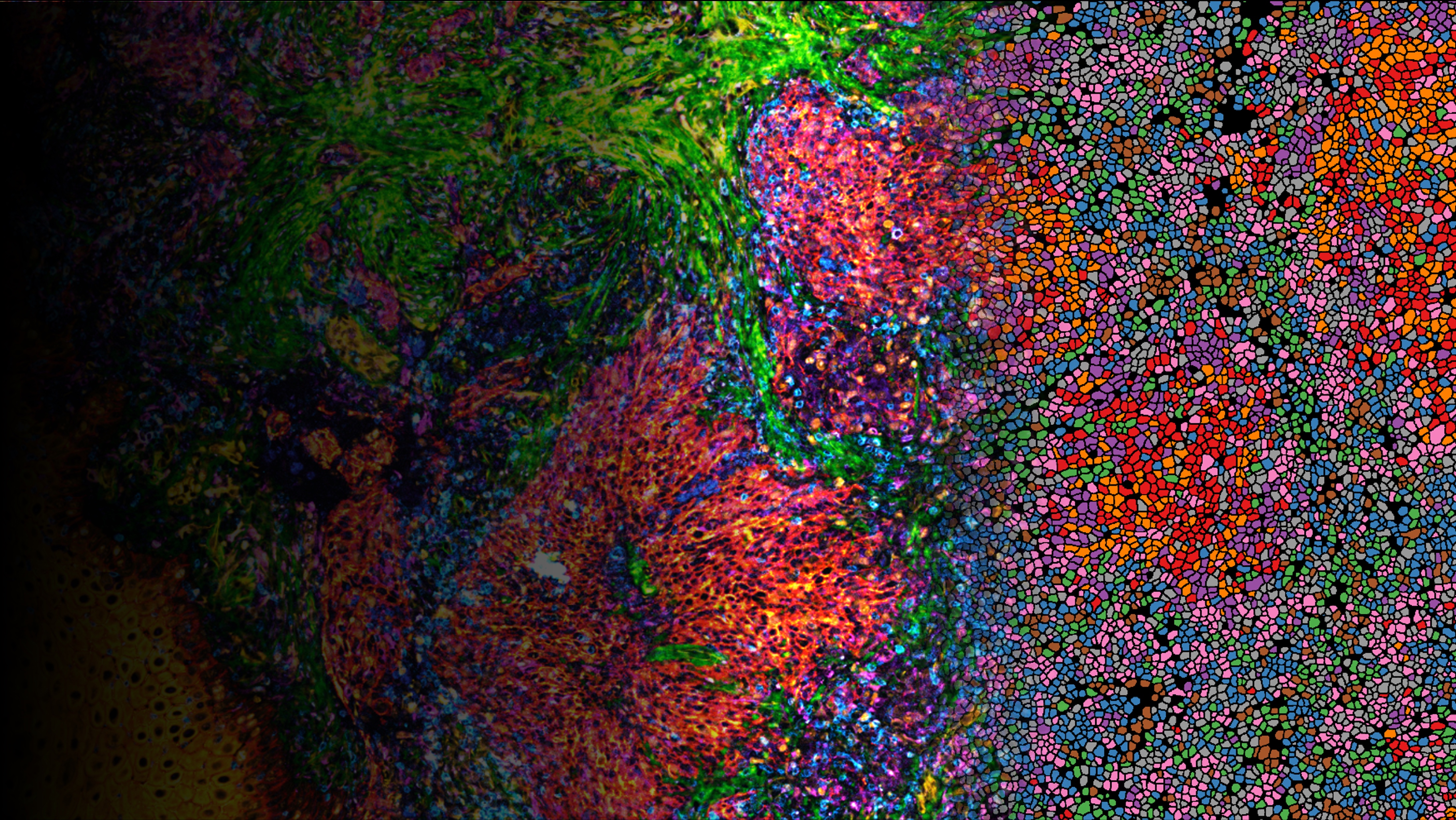
Multiplex immunofluorescence imaging of a head and neck cancer tissue sample. The left side shows the original microscopy image, which transitions into cell segmentation toward the right and that guides the deep visual proteomics sampling.
© Sonja Fritzsche, Coscia lab, Max Delbrück Center
Contacts
Dr. Fabian Coscia
Group Leader,
Spatial Proteomics
Max Delbrück Center
fabian.coscia@mdc-berlin.de
Gunjan Sinha
Editor, Communications
Max Delbrück Center
+49 30 9406 – 2118
Gunjan.Sinha@mdc-berlin.de or presse@mdc-berlin.de
- Max Delbrück Center
-
The Max Delbrück Center for Molecular Medicine in the Helmholtz Association lays the foundation for the medicine of tomorrow through our discoveries of today. At locations in Berlin-Buch, Berlin-Mitte, Heidelberg, and Mannheim, interdisciplinary teams investigate the complexity of disease at the systems level – from molecules and cells to organs and entire organisms. Together with academic, clinical, and industry partners, and as part of global networks, we turn biological insights into innovations for early detection, personalized therapies, and disease prevention. Founded in 1992, the Max Delbrück Center is home to a vibrant, international research community of around 1,800 people from over 70 countries. We are 90 percent funded by the German federal government and 10 percent by the state of Berlin.