Heart repair via neuroimmune crosstalk
Each year, more than 300,000 people in Germany have a myocardial infarction – the technical term for heart attack. The number of people surviving a heart attack has increased significantly, but this severe cardiac event causes irreparable damage to their hearts. A heart attack occurs when blood vessels that supply blood and oxygen to the heart muscle become blocked, causing part of the heart muscle tissue to die. This damage is permanent because the human heart has no ability to grow new heart muscle cells. Instead, connective tissue cells known as fibroblasts migrate into the damaged area of the heart muscle. They form scar tissue that weakens the pumping power of the heart. Previous attempts to use stem cells to treat infarction-damaged hearts have not been very successful.
This will help us understand why heart muscle tissue is unable to regenerate in humans.
The team led by Dr. Suphansa Sawamiphak, head of the Cardiovascular-Hematopoietic Interaction Lab at the Max Delbrück Center, is looking at the process from a different angle. “We know that both signals from the autonomic nervous system and the immune system play a pivotal role in scarring and regeneration,” says Sawamiphak. “So it stands to reason that the communication between the autonomic nervous and immune systems determines whether heart muscle scarring will occur or whether the heart muscle can recover.” It is also known that macrophages play a role in both processes. But how is this decision made?
To address this question, the researchers are studying zebrafish larvae. The fish can be easily modified and are also optically transparent, making internal processes easy to observe in the living organism. “Plus, they can fully regenerate their heart after an injury,” says Onur Apaydin, first author of the study published in “Developmental Cell.”
Signaling for regeneration
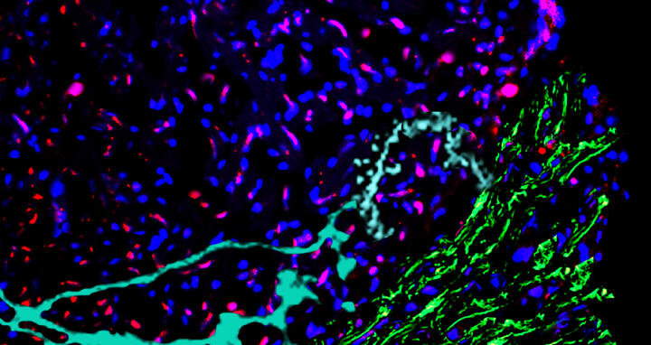
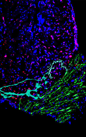
Cryoinjured section of a zebrafish heart: Immunofluorescence staining elucidates cellular and extracellular compositions pivotal for cardiac repair. All cell nuclei are seen in blue, while the red stain delineates cardiomyocytes. The extracellular matrix, crucial for structural integrity and signaling, is highlighted in green. Cyan staining reveals neurons, underscoring the neuro-cardiac interactions during regeneration.
The researchers used zebrafish larvae whose heart muscle cells produce a fluorescent substance, making it easy to detect them under a microscope. They then induced an injury similar to a myocardial infarction in the larval hearts and blocked several receptors on the surface of the macrophages. The result was that adrenergic signals from the autonomic nervous system determined whether the macrophages multiplied and migrated into the damaged site. These signals also played an important role in regenerating heart muscle tissue.
In the next step, the researchers engineered genetically modified zebrafish in which the adrenergic signal reached the macrophages but could not be transmitted from the receptor into the cell’s interior. “This showed that signal transmission is crucial for heart regeneration,” says Apaydin. If signaling is interrupted, the scarring process is triggered instead.
“Our findings indicate that this is a key regulator of crosstalk between the nervous and immune systems,” says Apaydin. When macrophages are activated by the adrenergic signals of the autonomic nervous system, they in turn communicate with fibroblasts. Fibroblasts that promote regeneration alter the extracellular matrix at the damaged site. This ultimately creates a microenvironment conducive to the growth of blood and lymph vessels and to the development of new heart vessels. If, on the other hand, the signal is blocked, fibroblasts infiltrate the site and cause scarring – similar to what occurs in the human heart after a heart attack.
“We next want to examine in detail how signaling differs between zebrafish and humans,” says Sawamiphak. “This will help us understand why heart muscle tissue is unable to regenerate in humans.” The team also hopes to identify potential targets for influencing the interaction between the nervous and immune systems in a way that promotes the regeneration of heart muscle tissue and the maintenance of heart function in heart attack patients.
Text: Stefanie Reinberger
Further information
Literature
Onur Apaydin et al. (2023): “Alpha-1 adrenergic signaling drives cardiac regeneration via extracellular matrix remodeling transcriptional program in zebrafish macrophages”. Developmental Cell, DOI: 10.1016/j.devcel.2023.09.011
Picture to download
Cryoinjured section of a zebrafish heart: Immunofluorescence staining elucidates cellular and extracellular compositions pivotal for cardiac repair. All cell nuclei are seen in blue, while the red stain delineates cardiomyocytes. The extracellular matrix, crucial for structural integrity and signaling, is highlighted in green. Cyan staining reveals neurons, underscoring the neuro-cardiac interactions during regeneration. Photo: Onur Apaydin, Max Delbrück Center
Contacts
Dr. Suphansa Sawamiphak
Head of the Lab “Cardiovascular-Hematopoietic Interaction”
Max Delbrück Center
Suphansa.Sawamiphak@mdc-berlin.de
Jana Schlütter
Editor, Communications Department
Max Delbrück Center
+49 30 9406 2121
jana.schluetter@mdc-berlin.de or presse@mdc-berlin.de
- Max Delbrück Center
-
The Max Delbrück Center for Molecular Medicine in the Helmholtz Association (Max Delbrück Center) is one of the world’s leading biomedical research institutions. Max Delbrück, a Berlin native, was a Nobel laureate and one of the founders of molecular biology. At the locations in Berlin-Buch and Mitte, researchers from some 70 countries study human biology – investigating the foundations of life from its most elementary building blocks to systems-wide mechanisms. By understanding what regulates or disrupts the dynamic equilibrium of a cell, an organ, or the entire body, we can prevent diseases, diagnose them earlier, and stop their progression with tailored therapies. Patients should be able to benefit as soon as possible from basic research discoveries. This is why the Max Delbrück Center supports spin-off creation and participates in collaborative networks. It works in close partnership with Charité – Universitätsmedizin Berlin in the jointly-run Experimental and Clinical Research Center (ECRC), the Berlin Institute of Health (BIH) at Charité, and the German Center for Cardiovascular Research (DZHK). Founded in 1992, the Max Delbrück Center today employs 1,800 people and is 90 percent funded by the German federal government and 10 percent by the State of Berlin.


