Stopping stomach cancer before it starts
Joint press release by Charité — Universitätsmedizin Berlin and the Max Delbrück Center
Disease almost never develops overnight. If illnesses are detected in later stages, however, they often prove difficult to treat — the same holds true for stomach cancer. Years before the condition sets in, the stomach lining gradually begins to change. The most frequent trigger: The common stomach bacterium Helicobacter pylori (H.pylori), which is primarily transmitted within families. If parents or grandparents have had stomach cancer caused by H.pylori infection, their descendants are also at high risk of developing the disease.
A team led by Dr. Michael Sigal, Professor of Translational Gastrointestinal Oncology at Charité – Universitätsmedizin Berlin and Group Leader of the Gastrointestinal Barrier, Regeneration and Carcinogenesis lab at the Berlin Institute of Medical Systems Biology of the Max Delbrück Center (MDC-BIMSB), and Dr. Manqiang Lin at Charité, has uncovered how exactly the bacteria remodel the stomach lining to promote cancer. “If we understand what happens in the stomach tissue even before cancer develops, we might be able to intervene,”explains Sigal. The research was published in “Nature Communications.”
Sigal is also a member of the recently launched Berlin Cluster of Excellence ImmunoPreCept, a collaboration spearheaded by researchers at Charité and MDC-BIMSB and that includes several other partners. ImmunoPreCept is dedicated to identifying disease-causing processes and halting them before it is too late. In this context, researchers from the cluster and other institutions have joined forces to, among other things, determine what occurs in the gastric mucosa at the cellular and molecular levels when it is exposed to H.pylori over extended periods of time.
The fact that H.pylori promotes the development of cancer has long been known. But what types of cells are involved? In what order do they relay signals? And which of these signals is the most important? “We knew from previous studies that the bacterium disturbs the balance of growth signals in the stomach lining. What we were missing was the link between the inflammation that had been triggered and tissue remodeling. This is because the mucous membrane does not behave the way one would actually expect. It grows rapidly, though without stem cells multiplying in the process. “We thought there must be another program at work that has been overlooked until now,” explains Sigal.
Stepwise like a game of chess
The researchers harnessed single-cell sequencing to analyze tens of thousands of cells from the diseased stomach lining and determined, for each cell type, which genes are turned on during an infection. In order to identify the cause and effect, they used animal models in which specific genes in certain cells were selectively inactivated. Tiny, laboratory-grown miniature versions of the gastric mucosa, known as organoids, and assembloids — combinations of mucosal and connective tissue cells developed specifically for this purpose — were used in subsequent investigations to simulate communication between the different cell types and to selectively interrupt this communication. In this way, the researchers were able to observe which cells in the tissue are actually located adjacent to one another and exchange signals. They then used publicly available datasets to verify whether these findings also apply to human tissue.
The research team encountered a program that works similar to a game of chess: Each step paves the way for the next. The chain culminates in precancerous lesions and then cancer, explains Dr. Giulia Beccaceci, first author of the study and an early-career researcher in Sigal’s lab. “The infected stomach lining doesn’t just grow faster, but actually transitions to a fundamentally different state. A process is gradually activated that normally occurs only during embryonic development and wound healing.”This is the reason why the tissue undergoes such lasting changes.
The process sets in when H.pylori overcomes the stomach’s natural defense system. A signal fails to be transmitted that normally ensures the controlled renewal of the surface cells of the mucous membrane and their tolerance of bacteria. Consequently, the immune system is alerted; immune cells migrate to the site and produce pro-inflammatory mediators, primarily interleukin-1β. This, in turn, does not affect the mucous membrane itself, but rather the underlying connective tissue. From there, the decisive signal is finally transmitted: A tissue hormone that normally helps heal injuries switches the mucous membrane into repair mode. As a result, the cells grow uncontrollably and divide more frequently. “So connective tissue isn’t just a bystander, but the actual switch,” Beccaceci adds.
Disease progression can be halted
Now that researchers better understand how H. pylori infection leads to chronic gastritis, precancerous lesions and ultimately cancer, they may be able to prevent disease progression. “People with an increased risk — for example, due to a family history of stomach cancer, symptoms, or a known infection — should be tested for the stomach bacterium and, if the test is positive, treated with antibiotics,” says Sigal. “This is simple and has been proven to reduce the risk of stomach cancer. This is due to the fact that it removes persistent irritation from the tissue.”
The gastric mucosa, however, does not fully return to normal after antibiotic treatment in all affected individuals. In some cases, the tissue has already been permanently reprogrammed, and the risk that cancer will develop remains. “In order to reliably identify these individuals, we are currently developing markers that can detect, in mucosal tissue samples, whether the tissue is on its way to becoming precancerous,” explains Sigal. “In addition, we now understand the individual steps in the chain and, as a result, the potential targets for drugs.” Consequently, this triangular communication via connective tissue could be disrupted by making these cells unresponsive to the inflammatory mediator interleukin-1β. That would prevent tissue changes and abnormal mucosal growth.”
Researchers are also encouraged by findings from previous population studies, which showed that regular use of known anti-inflammatory drugs was associated with a lower risk of gastrointestinal tumors. These medications also block a component of the pathway now identified: the enzyme COX‑2. This confirms that the mechanism works, and the new findings will help interrupt the pathway in an even more targeted manner and with fewer side effects.
About the study
In addition to scientists at Charité, MDC-BIMSB and the ImmunoPreCept Excellence Cluster, other collaborators included: The Institute for Experimental Internal Medicine at Otto von Guericke University Magdeburg, the Max Planck Institute for Infection Biology, Berlin, the Berlin Institute of Health (BIH) at Charité, the BIH Charité Clinician Scientist Program, and the Berlin School of Integrative Oncology (BSIO) at Charité. The research was funded by, among others, the European Research Council (ERC Starting Grant REVERT), the Deutsche Forschungsgemeinschaft, the Federal Ministry of Research, Technology, and Space, the Einstein Center 3R, and the ImmunoPreCept Excellence Cluster.
Text: Charité – Universitätsmedizin Berlin
Further information
ImmunoPreCept
MDC BISMB
How H. pylori alters tissue
Literature
Giulia Beccaceci, Stefanie Müllerke, Hilmar Berger, et al. (2026): “Helicobacter pylori triggers gastric mucosal remodeling toward a fetal-like transcriptional program via stromal IL-1β signaling.” Nat Coms, DOI: 10.1038/s41467-026 – 77520‑1
Contact
Dr. Michael Sigal
Group Leader
Gastrointestinal Barrier, Regeneration and Carcinogenesis lab
MDC-BIMSB
+49 30 450 614 055
michael.sigal@mdc-berlin.de
Gunjan Sinha
Editor, Communications
Max Delbrück Center
+49 30 9406 – 2118
presse@mdc-berlin.de
Image for download
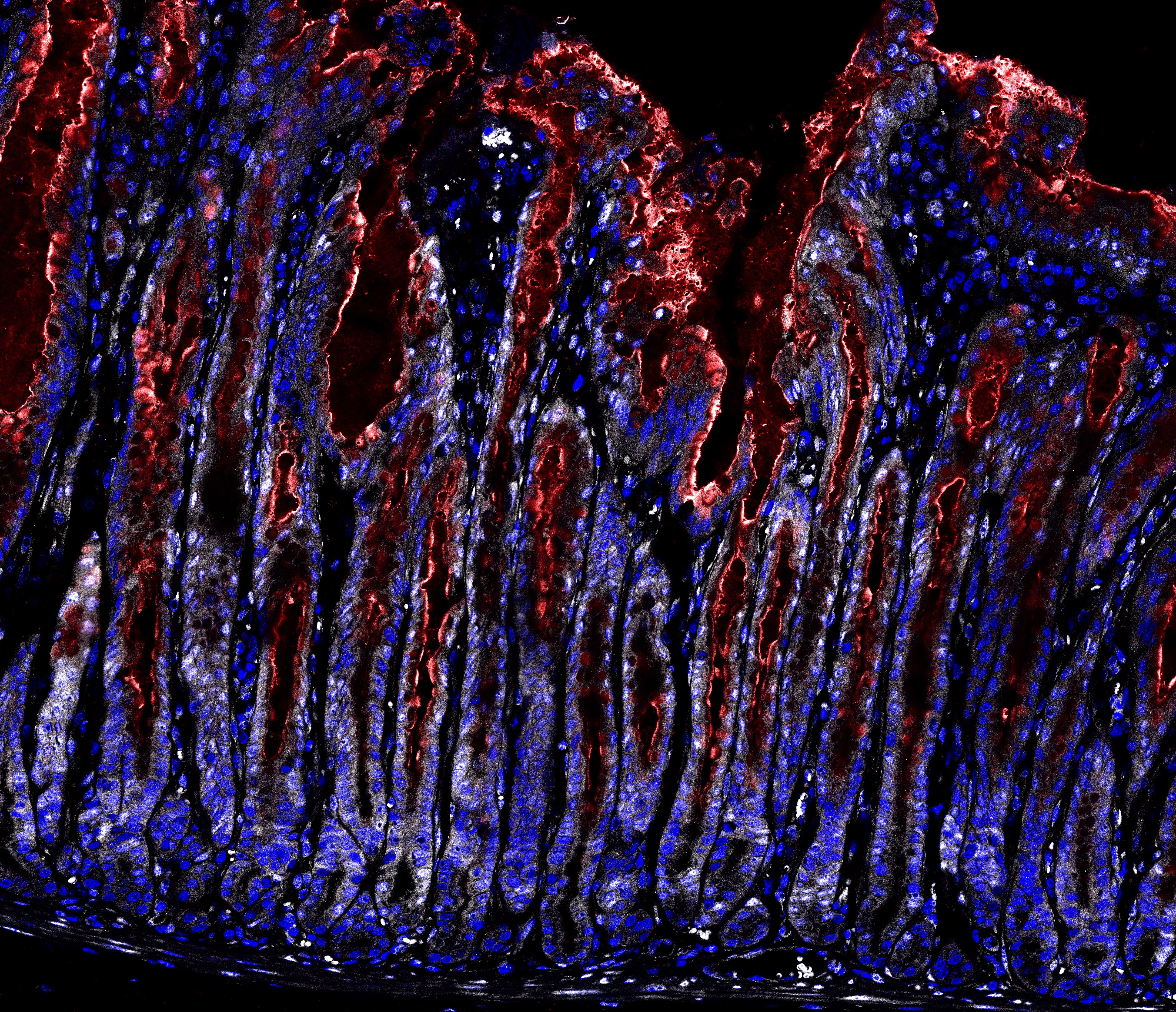
Caption: Stomach tissue infected with Helicobacter pylori. What starts off as a repair response, transitions into a chronic condition after years of infection and creates the preconditions for precancerous lesions. The mucus-producing surface cells are stained red. The active signaling pathway, which induces rapid tissue growth, is shown in white. In a healthy stomach, these processes are not active. © Charité | Michael Sigal
- Max Delbrück Center
The Max Delbrück Center for Molecular Medicine in the Helmholtz Association lays the foundation for the medicine of tomorrow through today’s discoveries. At locations in Berlin-Buch, Berlin-Mitte, Heidelberg, and Mannheim, interdisciplinary teams investigate the complexity of disease at the systems level – from molecules and cells to organs and entire organisms. Together with academic, clinical, and industry partners, and as part of global networks, we turn biological insights into innovations for early detection, personalized therapies, and disease prevention. Founded in 1992, the Max Delbrück Center is home to a vibrant, international research community of around 1,800 people from over 70 countries. We are 90 percent funded by the German federal government and 10 percent by the state of Berlin.